Pixel corrections for thick sensor detectors
C. López Jurado, S. Selleck, J. Just, M. Ramakrishnan, S. Ansell, C. Weninger, Z. Matěj
MAX IV Laboratory, Lund University, Sweden
zdenek.matej@maxiv.lu.se
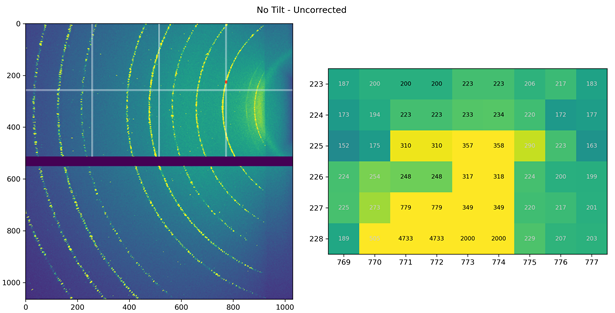
Flat hybrid pixel detectors present the most common instruments for recording photon counts in crystallographic experiments at accelerator-based light sources as well as in X-ray laboratories. To optimize efficiency of the detection process the ratio of detector sensor thickness and pixel face size is commonly set quite high. A narrow X-ray beam entering such a detector at an oblique angle is absorbed in multiple consecutive pixels. This is causing an effective shift of the detected signal known as the “parallax” effect. The absorption of X-rays in the detector sensor is also more complete. The latter is called an “oblique incidence effect”. These effects can cause a characteristic blur of diffraction spots depending on the detector incidence angle. Appropriate corrections are well established in software for single crystal diffraction data processing [1] and known in software for azimuthal integration of diffraction patterns from X-ray area detectors [2]. A framework [3] was created to simulate the parallax and oblique incidence effects by means of point spread function (PSF) using a simple raytracing of the interaction of X-rays with the detector and assuming a point X-ray source. The model can describe realistic detector cases including gaps between detector submodules knows as “wide pixels” (Figure 1). Detector PSF is represented by a sparse matrix and so the calculation of the blurred image for any type of ideal simulated signal is very effective. More complex is the deconvolution of known simulated PSF from a measured images that should result in position and intensity corrected diffraction images and optimally also sharpen diffraction spots. Several methods (Richardson-Lucy, Deep learning, and Direct inversion) are presented and compared. Their applications for diffraction data processing are briefly discussed.
1. W. Kabsch, X-ray Detector Software, XDS version: March 15, (2007), https://xds.mr.mpg.de/html_doc/Release_Notes.html (visited May 5th, 2023)
2. J. Kieffer et al., pyFAI documentation: Deconvolution of the Thickness effect, (2023), https://pyfai.readthedocs.io/en/master/usage/tutorial/ThickDetector/deconvolution.html (visited May 5th, 2023)
3. S. Selleck, C.L Jurado and Z. Matěj, xtrace: Removing Detector Depth Blur in Near Field Scattering - An Initial Exploration, (2023), https://github.com/maxiv-science/xtrace (visited May 5th, 2023)
|
|
|
Figure 1. Detector submodules are depicted in the upper left part. So called “wide-pixels” are visible in the enlarged image area on the right. |