Structural analysis of hard coatings and thin films at DEP FMPI CU in Bratislava
T. Roch, B. Grančič, M. Mikula, P. Ďurina, M. Gregor, T. Plecenik, L. Satrapinskyy
Department of Experimental Physics, Faculty of Mathematics, Physics and Informatics, Comenius University in Bratislava, Mlynska dolina F2, 842 48 Bratislava, Slovak Republic
roch@fmph.uniba.sk
Historically, X-ray structural analysis has been performed at the Comenius University in Bratislava since the 1950s. Basic research in the solid state physics and development of X-ray diffraction methods continued after establishing of Faculty of Mathematics and Physics by the separation of the physical and mathematical departments from the former Faculty of natural sciences in 1980.
The Laboratory of structural analysis at the Faculty of Mathematics, Physics and Informatics of Comenius University in Bratislava is today part of a larger complex of Laboratories of Advanced Technologies established in 2010 which is located at the Department of Experimental Physics. Our research group mainly focuses on three core topics: hard coatings for industrial applications and extreme environmental conditions, superconducting cryoelectronic heterostructures, development of novel simple gas sensors based on metal oxides. The investigated samples are in the form of thin layers, and the laboratory equipment fully enables their preparation and analysis.
Laboratories of Advanced Technologies contain set of PVD techniques like evaporation, magnetron sputtering (DC, RF), advanced sputtering methods (HiPIMS, HiTUS) and pusled laser deposition equipped with in-situ RHEED and ellipsometry. The micro- and nano-structures for test devices are fabricated using optical and e-beam lithography and by focused ion beam. Elemental chemical analysis is performed using energy- and wavelength-dispersive X-ray spectroscopy implemented in scanning electron microscopes. X-ray photoelectron spectroscopy provides extended chemical analysis. Electrical transport, magnetometry and thermal measurements can be performed using the multifunctional physical properties measurement system in temperature range of 50mK – 500K and magnetic field up to 14T. A complete characterization of mechanical and tribological properties is provided at the detached workplace of our group in Turany.
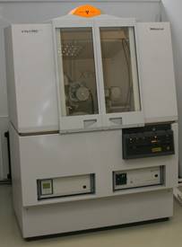
The main analytical facility for structural analysis is the diffractometer PANalytical X'pert Pro MRD (Fig.1), commissioned in October 2011. Within available budget the diffractometer was procured with options which could cover wide range of measurement modes of structural analyses of thin films. The diffractometer uses ceramic 1.8 kW X-ray tube (CuKa radiation) with long fine focus capable to flip into point focus. A precise motorized sample stage on the Eulerian cradle (X, Y, Z, azimuth, tilt) allows texture and stress analyses and also in-plane diffraction mode using an included option. Measurement setups are easily interchangeable using pre-aligned PreFIX optical modules. For high- resolution diffraction or (non-) specular reflectivity the parabolic X-ray mirror, 4-bounce Ge (022) Bartels monochromator with 3-bounce Ge (022) channel-cut analyzer, parallel plate collimator or variable detector slits are available. Monocapillary is suitable for texture, stress or in-plane diffraction measurements in combination with parallel plate collimator in front of detector. The diffractometer is routinely used for fast screening of deposited hard coatings. Typically, the comparison of coplanar grazing incidence diffraction together with fast symmetrical Bragg-Brentano scanning with PIXcel detector provides sufficient data for fast qualitative phase analysis and microstructure parameters estimation together with preferential orientation and stress-states. Short X-ray reflectivity measurements help in determination of mass density and thin films thickness. Although samples surface plane is positioned vertically, it is also possible to measure powders in Bragg-Brentano setup with appropriate measures.
Our research in hard coatings is focused on transition metal (TM) nitrides and borides. For example, TM diborides often contain vertically elongated nanocrystallites of the AlB2-type hexagonal phase embedded in a boron-rich amorphous tissue phase. For the more detailed structural analysis at nanoscale it is necessary to utilize e.g. transmission electron microscopy [1]. We have good cooperation with the Slovak Academy of Sciences or the Slovak Technical University in using their TEM equipment as an external service. Advantage is the ability to prepare our own lamellae using our focused ion beam equipment.
The mostly used monocrystalline silicon or sapphire substrates induce strong bi-axial texture especially in thin superconductor or metal oxide thin films [2,3]. Often the um-thick polycrystalline hard coatings show filamentary preferential orientation induced by deposition processes.
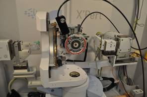
To investigate thermal stability, phase transitions and oxidation processes, the equipment allows in-situ measurements using a domed hot stage (AntonPaar DHS1100). In recent years, investigations of new hard coatings systems begin with theoretical calculations using density functional theory [4].
|
|
|
Figure 1. X’pert Pro MRD diffractometer. |
Figure 2. Goniometer with Eulerian cradle and attached HT chamber for in-situ measurements.
|
1. K. Viskupová, B. Grančič, T. Roch, Š. Nagy, L. Satrapinskyy, V. Šroba, M. Truchlý, J. Šilha, P. Kúš, M. Mikula, Scripta.Materialia, 229, (2023), 115365.
2. T. Roch, M. Gregor, P. Švec Jr., T. Plecenik, L. Satrapinskyy, M. Čaplovičová, R. Bystrický, P. Kúš, A. Plecenik, Apl. Surf. Sci.. 461, (2018), 233.
3. T. Roch, P. Durina, B. Grancic, M. Gregor, T. Plecenik, M. Truchly, M. Mikula, L. Satrapinskyy, P. Kus, A. Plecenik, Apl. Surf. Sci.. 312, (2014), 192.
4. T. Fiantok, V. Šroba, N. Koutná, V. Izai, T. Roch, M. Truchlý, M. Vidiš, L. Satrapinskyy, Š. Nagy, B. Grančič, P. Kúš, M. Mikula, J. Vac. Sci. Technol., A 40, (2022), 033414.
The work is the result of support under the Operational Program Integrated Infrastructure for the projects: Advancing University Capacity and Competence in Research, Development and Innovation (ACCORD) and UpScale of Comenius University Capacities and Competence in Research, Development and Innovation (USCCCORD, ITMS 2014+: 313021BUZ3), co-financed by the European Regional Development Fund.