Radon transformation in reciprocal space
M. Meduňa1, F. Isa2, F. Bressan3, H. von Känel2,3
1Department of Condensed Matter Physics, Masaryk University, Brno, Czech Republic
2Laboratory for Solid State Physics, ETH-Zürich, Zürich, Switzerland
3G-ray Switzerland SA Hauterive Switzerland
mjme@physics.muni.cz
Mapping intensity in reciprocal space around certain reciprocal lattice points is mostly realized in two-dimensions within the (QxQz) plane which is usually sufficient for any analysis of crystal lattice strain, mosaicity, structural quality, internal defect distribution or surface morphology and structure, depending on the scattering technique used. Nevertheless, in certain cases mapping of the scattered intensity in 3D space, out of the scattering plane e.g. along the Qy axis, is more and more demanded as well. Scanning reciprocal space in 3D however requires a well collimated x-ray beam in all directions, so that such experiments are realized mainly at synchrotron sources in order to have a sufficiently intense and parallel beam. Practically any 2D pixel detector is also extremely suitable for timely effective collection of reciprocal space maps (RSMs).
In this work we realize a collection of 3D RSMs using standard laboratory equipment with only a partially-collimated beam using a typical linear focus x-ray tube. We use a Rigaku SmartLab diffractometer equipped with a 2D HyPix area pixel detector for recording series of symmetric diffraction RSMs on microstructured semiconductor samples. The 2D detector is used in a linear mode for fast collection of typical 2D QxQz RSMs where the sample is scanned at many different azimuths with respect to the diffraction vector as an axis of rotation. The 3D intensity distribution in reciprocal space is then reconstructed by means of a Radon transform procedure, typically used for many decades in real space computer tomography (CT) [1]. The details of the technique used here in reciprocal space and experimental details realized can be found in recently presented work [2], including the details of the samples.
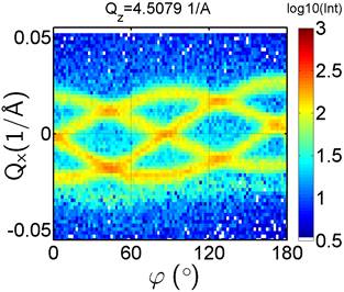
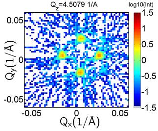
Figure 1. (left) Sinogram built from slices of typical QxQz RSMs for fixed Qz position close to SiGe (004) recorded at a series of azimuthal rotations of the sample containing four-fold symmetric diffraction superlattice satellites. (right) Reconstruction of the QxQy map in reciprocal space using the inverse Radon transform applied on the sinogram in the left panel can be understood as a given slice for fixed Qz through the 3D RSM. The four-fold structure of the four maxima is evident.
The method of applying the Radon transform for the reconstruction of RSMs is presented on Ge, GaAs and SiGe microcrystals epitaxially grown on patterned Si substrates [3]. Series of synchrotron experiments using a nanofocused beam, where 3D RSMs have already been measured on many samples, were previously presented in several of our publications [4,5,6], however in this study we compare similar laboratory measurements with the previously performed synchrotron experiments [2].
The QxQz RSMs obtained at various azimuths j are first decomposed into sinograms for all Qz positions, see an example for fixed Qz in the left panel of figure 1. This example is built from RSMs where certain (004) lateral diffraction satellites appear and their Qx position changes as the sample is rotated along j. After application of the inverse Radon transformation on this sinogam map, we can obtain the spatial distribution of these maxima within the QxQy plane perpendicular to the axis of rotation which shows four-fold symmetry, see the right panel of figure 1. Let us note that these satellites originate from a superlattice covering the faceted SiGe microcrystals. Comparing the new laboratory data [2] with previous synchrotron measurements [6], we get a very good agreement of the 3D spatial distribution of intensity.
1. P. Suetens, in Fundamentals of Medical Imaging, Cambridge University Press, 2009, pp. 35–41.
2. M. Meduňa, F. Isa, F. Brennan, H. von Känel, J. Appl. Cryst. 55, (2022) in print.
3. C.V. Falub, H. von Känel, F. Isa, R. Bergamaschini, A. Marzegalli, D. Chrastina, G. Isella, E. Müller, P. Niedermann, L. Miglio, Science, 335, (2012), 1330.
4. C. V. Falub, M. Meduňa, D. Chrastina, F. Isa, A. Marzegalli, T. Kreiliger, A.G. Taboada, G. Isella, L. Miglio, A. Dommann, and H. von Känel, Scientific Reports 3 (2013), 2276.
5. M. Meduňa, C.V. Falub, F. Isa, A. Marzegalli, D. Chrastina, G. Isella, L. Miglio, A. Dommann, and H. von Känel, J. Appl. Cryst, 49, (2016), 976.
6. M. Meduňa, C.V. Falub, F. Isa, D. Chrastina, T. Kreiliger, G. Isella, H. von Känel, J. Appl. Cryst., 47, (2014), 2030.
We acknowledge the staff of the ID01 beamline at the ESRF, Grenoble.