Non-traditional Applications of the Mössbauer Spectroscopy
A. Lančok
Institute of Inorganic Chemistry of the CAS, Husinec-Řež č.p. 1001, 250 68 Řež, Czech Republic
ada@iic.cas.cz
Nanomaterial is interesting material for artworks, biomedical and industrial applications especially in the field of nuclear installations. Our aim was to study of iron-containing nanoparticles in different type of materials (see Fig. 1); e.g. disks [1], powders [1-3], thin films etc. The methods for preparing of nanomaterials have attracted considerable scientific interest in recent years. These materials are structurally well ordered with very well-defined and exhibit unique physical and chemical properties determined by their practically applications. The aim of the lecture is to give an overview of current trends and perspectives in the research of above mentioned classes of nanomaterials.
Figure 1. Different type of nanomaterials.
Mössbauer spectrometry of the biological tissues confirms the presence of hematite, ferrihydrite and maghemite/magnetite in ferritin derived from human spleen and brain tissues. The minerals are present in a form of small (about 4-5 nm in size) grains with highly disordered structure. At room temperature all agglomerates of ferritin nanoparticles show non-magnetic behaviour. Employing Mössbauer effect measurements, the latter was determined to be of 16 K for the human spleen. Fig. 2 shows the evolution of Mössbauer spectra of low temperature dependencies.
|
|
|
Figure 2. Temperature dependences. |
Figure 3. Mössbauer spectra of ground natural vivianite crystals used for the model samples after exposition to a series of heating steps [2]. |
A study of pure synthetic (as well as partly oxidised) vivianite (materials for artworks) covering the whole extent of the temperature-related stability of its structure is being published for the first time in [2]. The results show that already temperatures around 70°C are damaging to vivianite, therefore, some of the recommended procedures during vivianite’s synthesis including digestion under increased temperatures should be avoided. Mössbauer spectra demonstrated that the temperature-induced oxidation of vivianite starts at 90°C, which corresponds with the increase of the amount of metavivianite. Fig. 3 shows the evolution of Mössbauer spectra of vivianite with increasing temperature, and illustrates the amounts of Fe2+ and Fe3+ after each heating step.
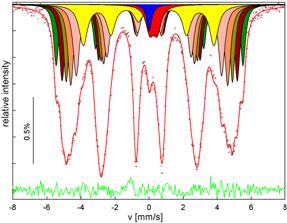
The transmission Mössbauer spectra of the powder and bulk samples show clear six-line pattern with only traces of a central single absorption line (shown in blue component in its central part). The six-line patterns in both spectra are assigned to magnetically ordered ferrite phase. They were fitted by 6 sextets. S1 through S5 represent those Fe atoms that have n Cr in their nearest shell with n = 0 up to 4. The sextet S6 with the lowest hyperfine field comes from the unresolved contribution of Fe atoms with 5 and more Cr nearest neighbours. Decomposition of the sextuplet part of spectra to components is plotted in Fig.4 in different shades of grey colour.
|
|
|
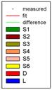
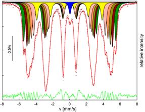
Figure 4. The decomposition of transmission Mössbauer spectra of the sample: (a) powder from of the surface of the disk and (b) bulk sample (transmission spectrum of the foil prepared from the disk).