Methods and tools for analysis of nanomaterials by means of atomic pair distribution function
Z. Matěj1,2, M. Dopita2, M. Paukov2, L. Havela2
1MAX IV Laboratory, Lund University, Lund, Sweden
2Faculty of Mathematics and Physics, Charles University in Prague, Praha, Czech Republic
zdenek.matej@maxiv.lu.se
Atomic Pair Distribution Function (PDF) describes distances between pairs of atoms in the matter on a nano-scale. A radial PDF weighted by atomic scattering factors can be obtained by means of X-ray and neutron total scattering on powder, nanocrystalline, amorphous or liquid samples. Beside Bragg intensity analysed by classical powder diffraction PDF brings information on a short range order from the diffuse scattering component. The number of scientific problems resolved by this method is rapidly growing thanks to multiple factors: as improved availability of dedicated neutron instruments, developments in X-ray detectors and laboratory instruments but also because of an advent of software and methods for straightforward PDF retrieval, modelling and analysis.
In this presentation we give a brief overview of the method and handy tools for PDF analysis. This is illustrated on an application case of X-ray data from nanocrystalline alloyed Uranium hydrides. We show some basic thoughts one shall do before starting a PDF experiment, list of some noticeable neutron instruments, demonstrate several tools for X-ray data reduction (see an example received from pyFAI [1] in Fig. 1 or srxplanar [2]) and a procedure to obtain the experimental PDF by means of PDFgetX3 [3]. We mention several tools for PDF simulations (DISCUS [4]) and data fitting (DiffPy suite [5]) that we considered very useful for working with nanocrystalline materials.
|
|
|
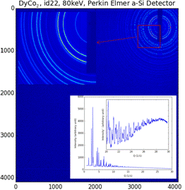
Figure 1. High energy synchrotron data from a temperature calibration sample of DyCo2. The inset shows image data reduced to a powder pattern using pyFAI. |
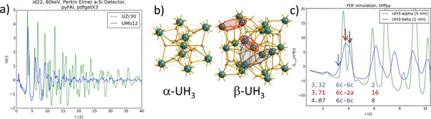
Our scientific problems is concerning U-hydrides. For us they present a probe to study an impact of expansion of the U lattice, allowing formation of U moments and their ferromagnetic ordering. Starting from (bcc) γ-U alloys using doping with Zr or Mo, different varieties of UH3 were synthtised [6]. Whereas Zr-dopping resulted in a fomation of (20 nm) nanocrystallites with a-UH3 cubic structure, pure Mo-alloyed UH3 is almost amorphous corresponding to very purely (1 nm) nanocrystalline material with b-UH3 like structure [7] (Fig. 2). Despite of these structural differences both types of hydrides behave similarly as ferromagnets with Curie temperature TC ≈ 170–200 K [6]. A striking phenomena observed in UH3 alloys is a large volume magnetostriction. This we tested in low temperature experiments.
|
|
|
Figure 2. (a) comparison of experimental PDF of
(UH3)0.70Zr0.30 and (UH3)0.88Mo0.12,
(b) unit cell structures of |
Analysis of real powder samples can be a complex problem especially if instrumental effects can influence the results and as the structure of the sample is often not ideal, e.g. becouse of presence of multiple phases or impurities. This is why beside fitting of PDF data with a structural model we employ alternative menthods of fitting of individual PDF maxima with phenomenological functions (a parSCAPE software can be useful for this [8]). Finally X-ray data analysis is complemented with preliminary neutron scattering simulations.
- G. Ashiotis, A. Deschildre, Z. Nawaz, J.P. Wright, D. Karkoulis, F.E. Picca, J. Kieffer, J. Appl. Cryst., 48, (2015), 510. doi: 10.1107/S1600576715004306
- X. Yang, P. Juhás, S.J.L. Billinge, J. Appl. Cryst., 47, (2014), 1273. doi: 10.1107/S1600576714010516
- P. Juhás, T. Davis, C.L. Farrow, S.J.L. Billinge, J. Appl. Cryst., 46, (2013), 550. doi: 10.1107/S0021889813005190
- Th. Proffen and R.B. Neder, J. Appl. Cryst., 30, (1997), 171. doi: 10.1107/S002188989600934X
- P. Juhas, C.L. Farrow, J. Liu, W. Zhou, P. Tian, Y. Shang, S.J.L. Billinge, Acta Cryst., A67, (2011), C143. doi: 10.1107/S0108767311096498
- I. Tkach, M. Paukov, D. Drozdenko, M. Cieslar, B. Vondráčková, Z. Matěj, D. Kriegner, A.V. Andreev, N.-T.H. Kim-Ngan, I. Turek, M. Diviš, L. Havela, Phys. Rev. B, 91, (2015), 115116. doi: 10.1103/PhysRevB.91.115116
- L. Havela, M. Paukov, I. Tkach, V. Buturlim, Z. Matej, S. Maskova, I. Turek, M. Divis, D. Drozdenko, M. Cieslar, M. Dopita, Z. Molcanova, M. Mihalik, MRS Advances, (2016). doi: 10.1557/adv.2016.287
- L. Granlund, S.J.L. Billinge, P.M. Duxbury, Acta Cryst., A71, (2015), 392. doi: 10.1107/S2053273315005276