MEASUREMENT
OF LATTICE PARTAMETERS OF SINGLE CRYSTALS AND THIN LAYERS
J. Drahokoupil1,*, Petr
Veřtát2, Kristina Richterová1, František
Laufek1
1Institue of Physics AS CR, Na Slovance 2, Prague 8, 182 21, Czech Republic
2 Faculty
of Nuclear Sciences and Physical Engineering CTU, Trojanova
13, Prague 2 120 00, Czech Republic.
*draho@fzu.cz
The precise measurement of lattice parameters plays an important role in determination a temperature of phase transitions or coefficients of thermal expansions. Usually it is performed on a powder or a bulk sample. Although the measurement of single crystal shows some complications, it has also many advantages. Firstly, the diffraction maximum of single crystal is narrower. Secondly, it can be usually measured to the higher diffraction angles because the Bragg peak overlap is not present there. Hence, the determination of d-spacing is more precise and consequently more precise lattice parameters can be obtained.
The following experimental setup was used: X´Pert PRO diffractometer with Co tube, parallel beam mirror in primary beam and parallel plate collimator (0,09°) in diffracted beam. The ATC-3 cradle equipped with the Peltier element was used for alignment of the sample and temperature controlling.
The measurements of lattice parameters on perfect Si single crystal, nice SmScO3 and TbScO3 crystals, non-ideal Ni-Mn-Ga crystals and SrTiO3 thin layer on DyScO3 substrate will be presented.
Data processing
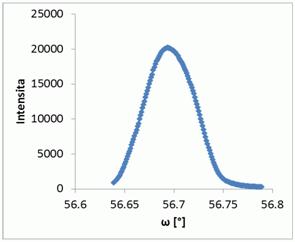
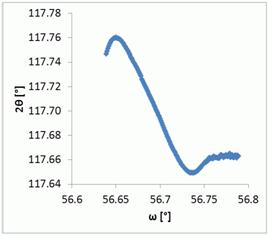
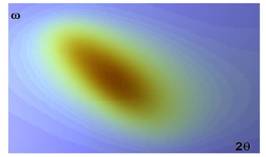
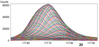
The diffraction maximum 333 of Si single crystal was carefully measured by two-axis scan with fine step in 2θ and ω, see Fig 1. Every 2θ-ω scan was fitted separately. The maximal intensity and its position of observed peak are presented in Fig. 2. It can be seen that around the maximum the position of peak depends linearly on offset (or ω). In order to reduce the measurement time, the exact position of diffraction maximum was obtained by simple extrapolation from two-axis scan with a coarser step in ω. In our case, we used parabolic extrapolation from three points around the scan with a maximal intensity.
The data processing was tested on the Si crystal. The 41 diffractions were measured, for every one 21 different 2θ-ω scans with a fixed offset. The lattice parameter was then refined using all 41 diffractions with an average error of 0,004° 2θ; the largest difference was 0,015° 2θ.
|
(a) |
(b) |
|
Figure 1. Diffraction 333 of Si single crystal. (a) top
view (b) side view with particular 2θ-ω
scan for fix offset. |
|
|
(a) |
(b) |
|
Figure
2. The properties of diffraction maximum: (a) maximal intensity of
particular 2θ-ω scan (b) its
position |
|