Structure analysis of nylon 6 nanofibers prepared by nanospider technology
A.Čajka1, P. Čapková1, J. Pavlík1, M. Munzarová2
1 Faculty of Science, J.E. Purkyně University, České mládeže 8, 40096 Ústí nad Labem
2 Nanovia, s.r.o. , Litvínov, Podkrušnohorská 271, 436 03 Litvínov-Chudeřín
Pavla.Capkova@ujep.cz
Polymer nanofibers find use in a wide range of areas such as: filtration [1], protective clothing, pharmaceuticals [2], tissue engineering [3], etc. The greatest attention of all polymeric fibers attracts nylon 6 due to its extraordinary properties: biodegradability, biocompatibility and good mechanical properties [4]. Various experimental arrangements and equipments have been described in literature for spinning of nylon 6 nanofibers either by melt spinning using extruder attached to a pump, see for example [5]) or by electrospinning with syringe attached to capillary tip connected to positive electrode [6]. Morphology and structure of the fibers prepared by these techniques has been characterized in dependence on spinning conditions and technology parameters [6,7]. However, the structure of fibers prepared by NANOSPIDER technology is less studied, although this technology is industrially used and for practice more significant. This study is devoted to the structure analysis of nylon 6 nanotextile prepared by NANOSPIDER technology under various conditions and for various technology parameters.
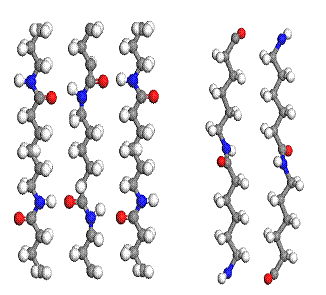
It is well known, that nylon 6 is polymorphic, having the following crystal structures: (1) The a- form described by Brill [8] and Holmes et al. [9] and (2) the g-form determined by Holmes et al. [9]. Both structures are monoclinic and differ from each other by arrangement of the polymeric chains, as one can see in figure 1. It is also known, that the melt spinning using extruder or electrospinning using capillary tip connected to positive electrode results in nylon 6 nanofibers composed of 3 structural phases: a, g and amorphous part and their mutual proportion depends on spinning parameters. Phase composition of nylon 6 nanofibers prepared by nanospider technology has not been investigated. Present work deals with the effect of spinning distance (i.e. distance between nanospider electrodes) on the structure of nylon 6 nanofibers, prepared in laboratory nanospider typ NS Production Line NS 1 WSU.
 Four nanotextile
samples have been prepared for spinning distance: 150, 200, 250 and 300 mm.
Wide angle X-ray scattering observable for all samples exhibits diffraction
profile, which indicates the presence of a and g phase with a contribution
of amorphous component. Example of such a profile is in the figure 2. Shoulders
on the profile indicate the presence of a-phase, while the
central main peak corresponds to g-phase. Large
broadening shows a very poor crystallinity and the presence of amorphous phase.
Four nanotextile
samples have been prepared for spinning distance: 150, 200, 250 and 300 mm.
Wide angle X-ray scattering observable for all samples exhibits diffraction
profile, which indicates the presence of a and g phase with a contribution
of amorphous component. Example of such a profile is in the figure 2. Shoulders
on the profile indicate the presence of a-phase, while the
central main peak corresponds to g-phase. Large
broadening shows a very poor crystallinity and the presence of amorphous phase.
Unraveling of the diffraction profiles for
all spinning distances used in our
experiment showed that:
- Increase of the spinning distance leads to higher degree of crystalinity, i.e. decrease of amorphous component and
increase of crystallite size
- Increase of the spinning distance led also to the increased
proportion of a-phase in the
sample.
- For all samples i.e. all spinning distances 150-300 mm, the nanotextile exhibits the strong texture, where the
polymer fiber axis in <020>
direction for both phases is preferentially oriented in the textile plane.
The degree of preferred orientation within individual fibers can`t be
estimated.
- Crystallite size roughly estimated was 5-10 nm, while the average
fiber diameter was 50-250 nm. Increasing spinning distance results in
thinner fibers and in better homogeneity in fiber diameters and smaller
number of failures having the shape of beads on fibers.
Figure
1: Arrangement of polymer chains in ab plane in crystal structure of a-phase (left) and g-phase
(right)

Figure 2: Diffraction profile of nylon 6 nanotextile
prepared for spinning distance 15 cm (radiation CuKa).
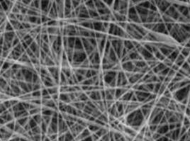
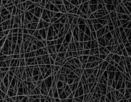
Figure 3: Nanotextiles prepared for
spinning distance 150 mm (left) and 300 mm (right).
[1] H. R. Ant M. P. Bajgai, Ch. Yi, R. Nirmala, K. T. Nam, W. Baek, H. Y. Kim., Colloids and Surfaces A: Physicochemical and Engineering Aspects. 370 (2010) 87-94.
[2] P. Raghavan, X. Zhao, J. K. Kim, J. Manuel, G. S. Chauhan, J. H. Ahn, C. Nan., Electrochim. Acta 54 (2008) 228-234.
[3] S. G. Kumbar, S. P. Nukavarapu, R. James, L. S. Nair, C. T. Laurencin, Biomaterials 29( 2008) 4100-4107.
[4] J. L. Pey, Anti-Corros. Methods Matter. 44 (1997) 94-99.
[5] T. D. Fornes, D.R. Paul, Polymer 44 (2003) 3945-3961.
[6] S. Zhang, W. S. Shim, J. Kim, Materials 30 (2009) 3659-3666.
[7] S. S. A. Deyab, M. H. E. Newehy, R. Nirmala, A. A. Megeed, H. Y. Kim, Korean Journal of Chemical Engineering. 30 (2013) 422-428.
[8] R. Brill, Z physik Chem B53(1943) 61-66.
[9] R. Holmes, R; C.W. Bunn, D.L. Smith, J Polym Sci 17 (1955) 619-624.