GISAXS application to a study of the nanoparticle self-assembly at the air-water interface
M. Jergel1, P. Šiffalovič1, K. Vegso1, M. Benkovičová1, E. Majková1,
K. Nygård2, O. Konovalov3
1Institute of Physics SAS, Dúbravská cesta 9, 845 11 Bratislava, Slovakia
2Dept. Chemistry & Mol. Biology, University Göteborg, 412 96 Göteborg, Sweden
3European Synchrotron Radiation Facility, BP 220, 38043 Grenoble, France
matej.jergel@savba.sk
Keywords: nanoparticles, GISAXS, Langmuir film
Abstract
An in-situ study of the colloidal silver nanoparticle self-assembly into a close-packed monolayer at the air/water interface followed by a 2D to 3D transition was performed by a fast tracking GISAXS technique in a Langmuir-Blodgett trough. Monitoring the immediate response of the system to the barrier movement enabled us to identify all relevant self-assembly stages including those far from the equilibrium. A new non-equilibrium phase before the monolayer collapse was found that is inaccessible by the competing direct space imaging techniques such as the scanning and transmission electron microscopies.
Introduction
Self-assembled arrays of chemically synthesized monodisperse nanoparticles are attractive for many novel and emerging applications. For example, the nanoparticle templates of plasmonic gold or silver nanoparticles provide a key to enhanced power conversion efficiency of future solar cells or may serve as unique substrates for the surface enhanced Raman scattering spectroscopy. The self-assembly takes place on the water surface in the Langmuir-Blodgett (LB) trough where a nanoparticle Langmuir film is formed and subsequently transferred onto a solid substrate. Macroscopic physical quantities such as the surface pressure, refractive index or surface potential of the nanoparticle Langmuir film are readily measureable while the conventional direct space imaging methods such as the scanning and transmission electron microscopy or nanoscale scanning probe techniques are inapplicable at the air/water interfaces due to the high vapour pressure and surface tension of the water subphase. Here, the reciprocal space techniques based on the scattering of X-rays or neutrons, in particular the synchrotron based grazing-incidence small-angle X-ray scattering (GISAXS), prove to be unique in revealing the atomic or molecular structure details at the air/water interface. We report here on the principal stages of a plasmonic nanoparticle Langmuir film compression at the air/water interface followed by a pressure release. The immediate film response to the surface pressure evolution is monitored in situ by a fast tracking GISAXS technique in order to detect transient phenomena far from the equilibrium. The structure evolution at nanoscale is related to the macroscopic behaviour monitored by the surface pressure, Brewster microscopy and imaging ellipsometry.
Experimental details
We studied Langmuir film composed of spherical Ag nanoparticles capped with oleic acid and oleylamine surfactant. The chemical synthesis was published elsewhere [1]. The surfactant prevents the nanoparticles from agglomeration. The diameter of the Ag nanoparticle core of 7±0.7 nm was determined by SAXS. The Ag nanoparticle solution in chloroform of 0.2 mg/ml concentration was spread on the deionised water subphase (specific electrical resistance >18 MΩ.cm) in a custom designed LB trough. After spreading the nanoparticles, the solvent was let to evaporate for 15 minutes before the measurement started. The in situ GISAXS measurements at the air/water interface during the barrier movement in the LB trough were performed at ID10B beamline at ERSF, Grenoble. The X-ray beam of the 300x100 mm2 size and 8 keV energy hit the air/water interface at 0.35º grazing angle. A fast 2D X-ray detector PILATUS 300K was used. The reciprocal space calibration was done by the silver behenate standard. The Brewster angle microscopy and null ellipsometry images were obtained by an optical microscope with digital output (Hitachi) and imaging ellipsometer (Accurion), respectively.
Results and discussion
To track in situ the evolution of the nanoparticle
order as a function of the surface pressure in the Langmuir trough during the
compression and expansion at a constant barrier speed of 26 cm2/min,
a continuous series of the GISAXS frames was recorded as a movie. The time between
the two successive frames of 1.87 s was short enough not to miss any
relaxation effect, hence, the immediate response was
monitored. The Fig. 1 shows the GISAXS patterns of the nanoparticle
Langmuir film before and after the compression. They consist of the nanoparticle interference function modulated by the nanoparticle form factor. The qz and qy are
the normal and lateral (in-plane) scattering vector components with respect to
the air/water interface. The 1st order Bragg rods suggest presence
of an ordered nanoparticle monolayer while the null ellipsometry image (Fig. 2) shows that it is
discontinuous at small surface pressure. Considering the hexagonally ordered
close-packed spherical nanoparticles, the lattice spacing before compression reads ![]() which for the first
Bragg rod maximum position
which for the first
Bragg rod maximum position ![]() =0.75 nm-1 gives the
nearest-neighbour interparticle
distance
=0.75 nm-1 gives the
nearest-neighbour interparticle
distance ![]() =9.7 nm. Assuming the mean nanoparticle
core diameter of 7 nm from SAXS, this gives some 1.4 nm for the
surfactant shell thickness. The
compression results in an intensity redistribution along qy of the central and 1st Bragg rods
(Fig. 1). On the former, the maximum intensity shifts from the critical
exit angle (Yoneda peak) to peak P1 (hidden by the specular beam stop) after
compression which indicates the nanoparticle layering. On the latter, peak P2
evolves at the position that suggests formation of a vertically correlated
second layer [2]. In particular, the AB stacking (hexagonal close-packed
bilayer) is manifested by the diffraction peaks P1(qy, qz)
and P2(qy, qz) located at (0, 2p/dz)
and (2p/d10, p/dz),
respectively, where the vertical lattice spacing dz is given as
=9.7 nm. Assuming the mean nanoparticle
core diameter of 7 nm from SAXS, this gives some 1.4 nm for the
surfactant shell thickness. The
compression results in an intensity redistribution along qy of the central and 1st Bragg rods
(Fig. 1). On the former, the maximum intensity shifts from the critical
exit angle (Yoneda peak) to peak P1 (hidden by the specular beam stop) after
compression which indicates the nanoparticle layering. On the latter, peak P2
evolves at the position that suggests formation of a vertically correlated
second layer [2]. In particular, the AB stacking (hexagonal close-packed
bilayer) is manifested by the diffraction peaks P1(qy, qz)
and P2(qy, qz) located at (0, 2p/dz)
and (2p/d10, p/dz),
respectively, where the vertical lattice spacing dz is given as ![]() . Considering
the in-plane interparticle distance D=9.7 nm, the calculated P2 peak position matches
perfectly that one observed in the GISAXS pattern.
. Considering
the in-plane interparticle distance D=9.7 nm, the calculated P2 peak position matches
perfectly that one observed in the GISAXS pattern.
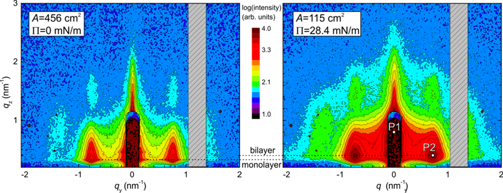
Figure 1. The GISAXS patterns of the nanoparticle
Langmuir film at the surface pressure 0 mN/m
(left) and 28.4 mN/m (right). The peaks P1 and P2
are associated with the AB stacked bilayer (see the
text). The dead area between
the detection modules of PILATUS 300K detector is hatched.
Based on the simultaneously measured pressure – area isotherm and GISAXS frames, we can identify four principal stages of the nanoparticle Langmuir film compression. In stage I, no measureable change in the surface pressure P is detected (Fig. 3a). We observe self-assembly of nanoparticles evidenced by the presence of Bragg rods in GISAXS (Fig. 1) which happens presumably within isolated self-assembled nanoparticle islands as the null ellipsometry shows (Fig. 2). The nanoparticle islands coalesce gradually into larger assemblies during the compression as indicated also by the Brewster microscopy (not shown). The surface elastic modulus E calculated from P derivative (Fig. 3a) has no physical meaning for the freely floating isolated islands here. The reduction of the Bragg rod width (Fig. 3c) is not observed because of a restricted size of the coherently scattering domains in the pristine islands controlled by the nanoparticle cumulative disorder. In stage II, we observe a steady increase in the surface pressure and elastic modulus
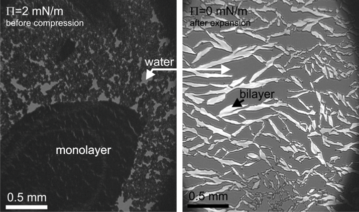
Figure 2. The null
ellipsometry images of the nanoparticle Langmuir film before the compression (left)
and after the expansion (right). The null imaging elipsometer was configured to
stop the polarized light reflected from the nanoparticle monolayer.
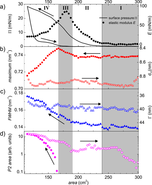
Figure 3. The major
compression stages of the nanoparticle Langmuir film. The arrows show the
compression and expansion periods. For a closer explanation see the text.
(Fig. 3a) as the proceeding island
coalescence gets gradually the larger assemblies into contact. This accumulates
stress at the assembly boundaries that is relieved by the nanoparticle
re-arrangements into a continuous close-packed monolayer. A nearly unchanged
Bragg rod maximum position and width (Fig. 3b,c)
suggest that the local hexagonal order from the original
islands is preserved. The nanoparticle
Langmuir film at the end of stage II is completely closed, being suitable for
the transfer onto a solid substrate to deposit a high-quality nanoparticle monolayer. In stage III, we observe a steeper
increase in the surface pressure followed by a maximum in the surface elastic
modulus which precedes a bilayer formation [3]. A
shift in the Bragg rod maximum position to the higher qy values suggests a
decrease in the lattice spacing d10
by approximately 0.1 nm (Fig. 3b) while a simultaneous increase in
the Bragg rod width (Fig. 3c) indicates a deterioration of the nanoparticle order due to the accumulated stress. Such a Bragg rod behaviour can be
explained by a slight compressive deformation of the polymer surfactant capping
the nanoparticles. This non-equilibrium compression
phase has never been observed in the experiments under steady-state conditions.
In stage IV, the peak P2 indicates a newly formed vertically correlated second nanoparticle layer allowing stress relief which results in
the reversed shift of the Bragg rod maximum position and increase in the d10 lattice spacing. The
Bragg rod width increases as well suggesting growing disorder. Such a simultaneous
growth of the lattice spacing and disorder is typical for the paracrystal model [4]. In the limit of a highly disorder paracrystal, the Bragg rod maximum position ![]() is controlled by the interparticle
distance D instead the lattice spacing d10,
i.e.
is controlled by the interparticle
distance D instead the lattice spacing d10,
i.e. ![]() instead
instead ![]() . The
. The ![]() at the
end of stage IV is in between that suggests a partial paracrystalline
disorder. The Bragg rod
maximum position and width are not recovered during the film expansion
suggesting a stable nanoparticle order in the bilayer. The drop in the P2 peak area may be explained by
decreasing amount of nanoparticles in the probed X-ray volume during the film
expansion when the bilayer islands are formed and go away from each other,
leaving free subphase surface behind (see also Fig. 2).
at the
end of stage IV is in between that suggests a partial paracrystalline
disorder. The Bragg rod
maximum position and width are not recovered during the film expansion
suggesting a stable nanoparticle order in the bilayer. The drop in the P2 peak area may be explained by
decreasing amount of nanoparticles in the probed X-ray volume during the film
expansion when the bilayer islands are formed and go away from each other,
leaving free subphase surface behind (see also Fig. 2).
Conclusions
We identified principal stages of the Ag nanoparticle Langmuir film formation using the fast in-situ GISAXS technique supported by Brewster angle microscopy and imaging ellipsometry. The nanoparticle monolayer formation takes place via coalescence of free self-assembled nanoparticle islands with the hexagonal close-packed order that persists in the coalesced assemblies up to a temporary squeezing of the lattice shortly before the monolayer. This transient phase has not been observed under steady-state conditions. The monolayer collapse takes place by flipping up the nanoparticles and the second layer formation with the AB-like crystallographic stacking and enhanced paracrystalline-like disorder. The Langmuir film expansion runs irreversibly by decomposition into bilayer islands without observable changes in the nanoparticle order inside, leaving free subphase surface behind.
References
1. K. Vegso, P. Šiffalovič, M. Weis, M. Jergel, M. Benkovičová, E. Majková, L. Chitu, Y. Halahovets, Š. Luby, I. Capek & A. Šatka, Phys. Stat. Sol., A 208, (2011), 2629-2634.
2. M. Fukuto; R. K. Heilmann, P. S. Pershan, A. Badia & R. B. Lennox, J. Chem. Phys., 120, (2004), 3446-3459.
3. S. Kubowicz, M. A. Hartmann, J. Daillant, M. K. Sanyal, V. V. Agrawal, C. Blot, O. Konovalov & H. Möhwald, Langmuir, 25. (2008), 952-958.
4. R. Lazzari, R., in X-ray and Neutron Reflectivity, edited by J. Daillant & A. Gibaud (Berlin-Heidelberg: Springer), 2009, pp 283-342.
Acknowledgement
This work was done during implementation of the project Research and Development Centre for Advanced X-ray Technologies, ITMS code 26220220170, supported by the Research and Development Operational Programme funded by the ERDF.