Simulation of reciprocal space maps from elastic
strain field in periodical nanostructures
L. Horak,
J. Matejova
Department
of Condensed Matter Physics, Faculty of Mathematics and Physics, Charles
University in Prague, Ke Karlovu
5, 121 16 Praha, Czech Republic
horak@karlov.mff.cuni.cz
Laterally periodical nanostructures epitaxially grown on substrates, such as quantum dot arrays
and wires, are extensively used in electronic and optoelectronic applications.
The strain field in the nanostructure, caused by an epitaxial mismatch, strongly
affects the material properties, e.g. electronic band structure or magnetic
anisotropy. This strain field can be investigated by means of High Resolution
X-ray diffraction (HRXRD).
The strain induced shifts of the atoms from
their bulk-lattice positions together with the shape of the objects are
manifested in the distribution of the diffracted intensity in the reciprocal
space in the vicinity of the Bragg diffraction maxima. The interpretation of
measured reciprocal space maps is not direct, the
strain field has to be determined by a comparison of the measured data and the
numerical simulation of the diffraction experiment. The shape of the objects
can be obtained by complementary methods, e.g. scanning electron microscopy
(SEM), and this known information is usually included in the model for the
computation of the strain field.
We will present the experimental technique
(coplanar HRXRD) for the measurement of reciprocal space maps, which can be
performed with a standard laboratory high-resolution diffractometer.
The simulation of the diffraction maps is based on the simple kinematic x-ray
scattering theory, which transforms the problem to the numerical computation of
Fourier-like integrals producing numerical difficulties to be treated.
The strain field entering into the
computation of the diffraction maps can be constructed in several ways
depending on specified problem we want to solve. From many of them, three
particular cases will be presented:
Firstly, one can be interested in the shape
of crystalline core of the nanostructure, while this information is not
accessible by SEM. If the composition of the nanostructure material is
homogenous and the elastic properties are known, the strain field can be
calculated using theory of elasticity by means of finite-element method (FEM). The
strain field is fully defined just by the object-core shape, which has to be
optimized.
Secondly, the nanostructure of a known shape
has inhomogeneous composition, which locally determines the unstrained lattice
parameter. The unknown distribution of occupancy enters into the strain
computation. The resulting strain field is given by the solution of elasticity
equations solved be FEM. If we have a model predicting the occupancy
distribution, e.g. based on atomic diffusion, we can optimize its parameters to
find agreement with the measured data.
Thirdly, the periodical nanostructures, made
of identical material to the substrate, are covered by an additional film,
which induces the strain in the objects. The task is to evaluate just the
strain field in the nanostructures, although there is no reliable model for
computation via the theory of elasticity. The strain field is parameterized and
directly optimized to get agreement of the experimental and simulated
diffraction maps.
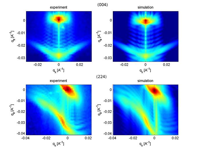
Figure 1: Reciprocal space maps near Bragg maxima
(004) and (224) measured and simulated in order to determine the shape of the (Ga,Mn)As micro-wires on GaAs substrate. The most intensive point is the substrate
peak of unstrained GaAs, the intensity distributed around
this peak is related to the strained parts of the substrate in the vicinity of
the wires. The intensity from partially relaxed (Ga,Mn)As wires is concentrated in the intense streak
bellow the substrate peak, the fringes in between are the thickness
oscillations from the wires.
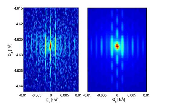
Figure
2: Measured (left) and simulated (right) diffraction map from oxidized Si-wires
for Ge nanoheteroepitaxy. The
intensity is present only along the truncation rods as a consequence of the lateral
periodicity of the wires. The lateral period is very small in this case, therefore the distance of the truncation rods in the
reciprocal space is large enough to see the individual satellites.