Investigations of real structure depth
distributions in metals
using diffraction techniques
Z. Pala
Department
of Solid State Engineering, Faculty of Nuclear Sciences and Physical
Engineering, Czech Technical University in Prague, Trojanova
13, 120 00 Prague 2, Czech Republic
zdenek.pala@fjfi.cvut.cz
In the case
of metals, X-ray diffraction (XRD) is often regarded as a surface technique
which reveals structural information about diffracting body only several
micrometers or tens of micrometers in thickness. Yet in comparison with
electron diffraction, assumption about surface sensitivity would be hardly
tenable. On the other end of the penetration scale, neutron diffraction is
firmly anchored because the neutrons penetrate metallic materials more easily
and the resulting data can be gained from depths amounting to centimetres.
Notion of penetration depth, which was
initially introduced by Cullity [1], is of vital
importance from the real structure point of view. The main reason is that metal
objects, especially those with surface treatment history, often show
significant depth gradients of macroscopic and microscopic residual stress,
grain sizes, or even texture. Gradients of these real structure characteristics
have direct impact on the object behaviour in real environment, affecting its
corrosion resistance, dynamic load stability, crack initiation and propagation
and other processes that can contribute to either prolongation or appreciable
shortening of fatigue life [2].
The effective penetration depth is
given by (i) absorption µ of the given volume for the impinging X-ray beam wavelength, (ii)
Bragg angle θ and (iii)
geometric alignment of the goniometer f(θ,ψ). Applying the well known Lambert-Beer
law for absorption, the ratio of intensities diffracted by a layer of thickness
dT
on the very surface and in the depth T
will be
 . (1)
. (1)
Upon
setting this ratio to a convenient constant of 1/e and taking a logarithm of
the (1), the term for effective penetration depth Te is calculated:
 . (2)
. (2)
Hence, Te determines the thickness
of a layer providing 63.2 % out of the entire diffracted intensity. Rigorously
said, the structural information gained from classical Bragg-Brentano goniometer or even grazing incidence diffraction changes
with the changing 2θ angle and
is, therefore, influenced by possible steep structural gradients. However, when
the gradients are not to be expected within the comparatively small thickness
comparable with Te, the
depth profiling can be performed with combining the chosen XRD technique on a
conventional laboratory diffractometers and
subsequent layer removal. Inherently destructive layer removal should be done
with minimal impact to the structure of the remaining layers; the most widely
used technique is electro-chemical polishing [3]. Even the laboratory diffractometers offer possibility to change X-ray tube and,
accordingly, the used wavelength. This would allow corresponding alteration of Te, yet given the variety of
X-ray tube selection, the scope of Te
for given metal is quite limited.
Another possibility for depth
distribution investigation in metals is to employ synchrotron radiation. Not
only is it tuneable with maximal energies peaking at 150 keV,
but the intensities increase manyfold. The
experimental set-up of 1ID C beamline at APS (Advanced Photon Source) in Argonne, USA
depicted in Fig. 1 is almost an ideal tool for defined depth profiling. The
incoming X-ray beam can be focused down to 2×5 μm2 and
with maximal energy of 130 keV can easily penetrate 1
cm of steel, with large detector array High Energy Detector Array or HYDRA© the
minimal acceptable transmission reaches 0.1 %.
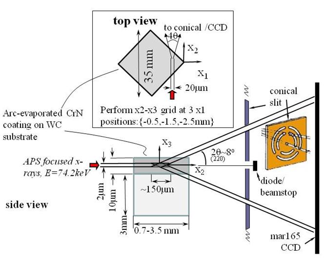
Figure 1. Experimental set-up of 1 ID C in APS, Argonne; courtesy of Jonathan Almer.
The presentation will deal with detailed
description of advantages and pitfalls of diffraction experiment at 1 ID C. Furthermore, a mutual comparison
between conventional XRD laboratory and synchrotron experiment results will be
offered. Tangibly, macroscopic residual stress profiles in ground steel will be
compared. Moreover, depth distributions of structure in plasma sprayed tungsten
and copper layers used as plasma facing components in tokamaks
obtained with use of neutron diffraction [4]and synchrotron radiation will be
discussed.
Possibility to measure and study depth
distributions of real structures parameters represent a further step in the
progress of diffraction techniques and bring them nearer to the wider public.
In this case, the industrial applications are, indeed, numerous and
significantly broaden horizons of material scientists and physical engineers
during new material evolution and manufacturing of metal objects used in
hi-tech applications.
1. B.D. Cullity, Elements of X-ray diffraction. Reading:
Addison -Wesley. 1956.
2. A. G. Youtsos, Residual stress and its effect on fracture
and fatigue. Springer 2006.
3. S. Lee, Y. Lee, M. Chung, Metal removal rate of the electrochemical
mechanical polishing technology for stainless steel – the electrochemical
characteristics, IMechE Vol. 220, 2006.
4. V. Luzin, J. Matejicek, T. Gnaupel-Herold, Through-thickness Residual Stress
Measurement by Neutron
Diffraction in Cu+W
Plasma Spray Coatings,
Materials Science Forum 652 (2010) 50-56.
The
research was supported by the Project MSM 6840770021 of the Ministry of
Education, Youth and Sports of the Czech Republic and by the Project
SGS10/300/OHK4/3T/14 of the Czech Technical University in Prague.