Periodic modulation of strain
fields and magentic anisotropy in (Ga,Mn)As/InAs/GaAs structures
T. Čechal1,
X. Martí1, L. Horák1, V. Novák2, K. Hruška2,
Z. Výborný2, T. Jungwirth2,3, V. Holý1
1 Department
of Condensed Matter Physics, Faculty of Mathematics and Physics, Charles
University, Ke Karlovu 5, 121 16 Prague 2, Czech Republic
2 Institute
of Physics ASCR, v.v.i., Cukrovarnická 10, 162 53 Prague 6, Czech Republic
3 School
of Physics and Astronomy, University of Nottingham, Nottingham NG7 2RD, United
Kingdom
cechal@mag.mff.cuni.cz
Thin layers of (Ga,Mn)As magnetic
semiconductor exhibit magnetic anisotropy which is strongly influenced by
lattice-matching strains introduced into these layers during epitaxial growth.
Laterally homogeneous strains can be induced by growing these layers on top of
GaAs (compressive strain) or (In,Ga)As (tensile strain) buffers [1].
Lithographic techniques can be used to create complicated strain patterns
leading to spatially varying magnetic anisotropy [2-4]. We combined e-beam lithography and dry etching
with molecular beam epitaxy to create ordered fields of InAs quantum dost on
GaAs(001) substrate which were subsequently
covered by a Ga0.95Mn0.05As capping layer.
High-resolution x-ray diffraction reciprocal-space mapping is
conventionally used to explore the strains in similar cases. However, the low
growth temperature required to incorporate Mn atoms into (Ga,Mn)As layers
causes that the crystal quality of such heterostructures is often not as good
as in the case of continuous epitaxial layers and previously reported
approaches to characterize the strain fields using the x-ray data are therefore
not directly applicable. Further complications arise from the combined effects
of strain and chemical roughness. Here we report on a simple fitting-free
methodology to evidence the presence of periodic strain fields from the
measured x-ray data and show how kinematical theory of x-ray scattering coupled
with the solution of elasticity equations can be used to determine the main
features of these strain fields.
The proceedings are organized as follows: first we present the
calculations of the strain fields in the case of (Ga,Mn)As layers grown on top
of periodic arrays of InAs quantum dots. Then we discuss the evaluation of the
intensity profiles along the satellites stemming from lateral periodicity and
we show that the presence of strain can be evidenced from the comparison of the
relative intensity of the first pair of satellites. Finally, we present the results
of x-ray diffraction experiment.
The strain in
(Ga,Mn)As/InAs/GaAs structures originates in different intrinsic lattice
parameters of the constituent materials. We used a combination of
Fourier and finite element methods to solve the equations of elasticity and
calculated the strain fields for a simplified model of the sample structure. In
this model we assumed that (1) the dot array is infinite and perfectly periodic
in the lateral direction, (2) the realistic dot shape can be approximated by a
truncated cone or pyramid, (3) elastic constants and lattice parameter of GaAs
can be used also for (Ga,Mn)As and (4) the surface is ideally flat and
force-free. A typical result of such a simulation is shown in Fig. 2 for conical dots with 40nm bottom radius, 10nm
top radius and 10nm high; the separation between dots is 100nm. We see that
tensile strain is confined to the vicinity of the dot apex whereas the regions
between the dots are compressively strained.
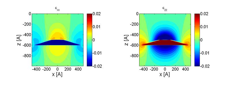
Figure 1.
Calculated exx and ezz
components of the strain tensor in a sample containing periodic array of
quantum dots. The dots have the shape of a 10nm high truncated cone with 40nm
bottom radius and 10nm top radius and are assumed to be composed of pure InAs.
The reciprocal-space distribution of measured intensity in an x-ray
scattering experiment exhibits lateral satellites stemming from the periodicity
of both the chemical composition and the strain fields. The intensity
distribution along a given satellite can be calculated within the framework of
kinematical theory of x-ray scattering and the resulting formula predicts that the
intensity ratio of the left and corresponding right satellite is equal to one
regardless of the diffraction if there is no strain in the sample; however, if
strain is present, this ratio is different from one in some diffractions as can
be seen in Fig. 2 which shows the calculated intensity distribution along the first
pair of satellites. The predicted asymmetry is indeed observed in the measured
x-ray data for the (224) diffraction (see Fig. 3) which provides a strong proof
of the presence of periodically modulated strain in the structures studied.
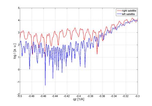
Figure 2. Intensity
distribution along the first pair of satellites near the (224) diffraction of
InAs calculated within the framework of kinematical scattering theory. The
asymmetry of the intensity profiles originates from epitaxial strains present
in the sample.
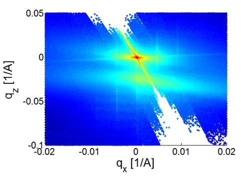
Figure 1. Measured
x-ray diffraction data close to the (224) substrate peak. The large area of
diffuse scattering corresponds to the (Ga,Mn)As layer; lateral satellites
originate from the periodicity of the underlying quantum dot array. The strong
asymmetry is a hallmark of the epitaxial strain.
References
1. M. Glunk, J. Daeubler, L. Dreher, S. Schwaiger,
W. Schoch, R. Sauer, W. Limmer, A. Brandlmaier, S. T. B. Goennenwein, C. Bihler
and M. S. Brandt, Phys. Rev. B, 79,
(2009), 195206.
2. J. Wunderlich, A.
C. Irvine, J. Zemen, V. Holý, A. W.
Rushforth, E. De Ranieri, U. Rana, K. Výborný, J. Sinova, C. T. Foxon, R. P.
Campion, D. A. Williams, B. L. Gallagher and T. Jungwirth, Phys. Rev. B, 76, (2007), 054424.
3. J. Wenisch, C.
Gould, L. Ebel, J. Storz, K. Pappert, M. J. Schmidt, K. Brunner and L. W.
Molenkamp, Phys. Rev. Lett., 99,
(2007), 077201.
4. T. Jungwirth, J.
Sinova, J. Mašek, J. Kučera and A. H. MacDonald, Rev. Mod. Phys,. 78,
(2006), 809.
Acknowledgements.
This work has been supported
by the European Commission projects NAMASTE (No.214499) and SemiSpinNet (No.
215368) and by the Academy of Sciences and Ministry of Education of the Czech
Republic (No. AV0Z10100521, No. KAN400100652, No. LC510, Praemium Academiae).
We acknowledge the staff at the ID10B beamline (ESRF), B. Bittová and J. Pospíšil (Charles University in Prague) for AFM and SEM
images and O. Caha (Masaryk University in Brno) for useful discussions.