Standing-wave-grazing-incidence x-ray diffraction from polycrystalline
multilayers
J. Krčmář1,
V. Holý2,
11Institute
of Condensed Matter Physics,
2Department of Physics of Electronic Structures,
3Institute
of Electrical Engineering Slovak Academy of Sciences, Dúbravská cesta 9, 84239 Bratislava,
krcmar@physics.muni.cz
In work [1] authors presented a new diffraction
method, which uses the concept of x-ray standing wave in the grazing-incidence
geometry, in which both the incidence and exit angles ai,f are close to the critical angle ac of total external reflection. If
the angle of incidence ai of the primary x-ray wave onto a periodic multilayer is close to ai or to a “Bragg-like” maximum on
the reflectivity curve, the interference of the transmitted and reflected waves
creates a standing wave pattern in the multilayer volume, the period of which
equals the multilayer period. If the multilayer contains crystalline grains
(crystallites), the intensity of diffraction from these crystallites depends on
the mutual position of the crystallites and the antinodes of the standing wave.
Similarly, the wave diffracted by the crystallites is reflected from the
multilayer interfaces, which results in a standing wave pattern as well. The
standing wave pattern is shifted by changing ai or af so that from the measured
dependence of the diffracted intensity on ai,f it is possible to determine the position of the diffracting
crystallites. Moreover, measuring the dependences of the diffracted intensity
on the in-plane scattering angle 2q at
various ai it is possible to determine the lateral sizes of the crystallites
in different depths in the multilayer. The theoretical description of the
scattering process is based on the Distorted-Wave Born approximation.
In Ref. [2], we proved the feasibility of this concept by measuring the standing-wave effects in C/Ni3N multilayers using synchrotron radiation, and we were able to determine the sizes of the crystallites in different depths in the multilayer and the thicknesses of amorphous Ni3N transition layers at the C/Ni3N interfaces.
In Ref. [3], we demonstrate that this
method can be carried out also at a modified x-ray diffractometer (Figure (1)),
and it is capable of studying the structure of crystalline layers in a periodic
multilayer at a laboratory (Figure (2)). We have used this method for the
investigation of the structure of Nb/Si multilayers, and we obtained the
thicknesses of amorphous and crystalline parts of the Nb layers. These
thicknesses agree very well with the transmission electron microscopy
observations.
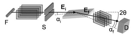
Figure 1. The geometry of the experimental method in the laboratory. X-ray tube has line focus F parallel to the surface plane. The incident Ei beam is restricted by a parallel plate collimator and slit S. The scattered beam Ef is restricted by a second parallel plate collimator. 2q is the scattering angle and ai and af are the incident and exit angles with respect to the sample surface.
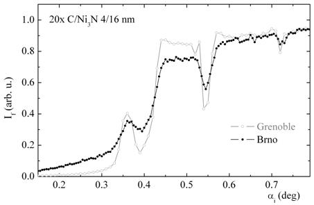
Figure 2.
Diffraction measured in the laboratory (
From the fits, it follows that the Nb layer consists in an amorphous part of the thickness of 2.0±0.5 nm lying at the Si/Nb interface (i.e., in the bottom part of the Nb layer) and of the crystalline part in the upper part of the layer at the Nb/Si interface. Figure (3) shows the comparison of the measured and fitted intensity curves. To demonstrate the sensitivity of the method, in Fig. (3) we have also plotted the calculated diffraction curves assuming that whole Nb layers are crystalline (dashed lines). From the figure, it is obvious that the agreement of these curves with the experimental data is much worse and the method is capable indeed to measure the thickness of the amorphous part of the Nb layer.
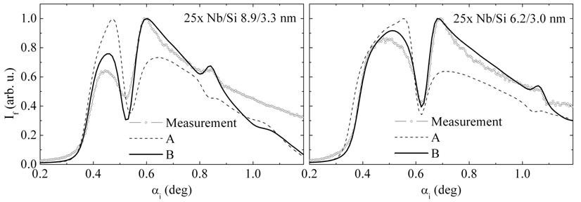
Figure 3. Measured (points) and fitted (lines and dash line) diffraction intensities. Curve A: all volume of Nb layers is crystalline. Curve B: bottom 2 nm thick parts of the Nb layers are amorphous.
Presented method is very suitable for a detailed study of sizes and positions of crystallites in periodic multilayers, as well as for the determination of the depth profile of the crystallite size.
References
1. P.F. Fewster, N.L. Andrew, V. Holý, K. Barmak, Phys. Rev. B., 72, (2005) 174105.
2. J. Krčmář, V. Holý, L. Horák, T.H. Metzger,
J. Sobota, J. Appl. Phys., 103, (2008) 033504.
3. J.
Krčmář, V. Holý,