Crystallization study of high plants photosystem II and chlorosomal
bacteriochlorophyll c aggregates
Tatyana Prudnikovaa, José A. Gavirac, Pavlína Řezáčovád, Michal Kutýa, b, František Váchaa, e, Jakub Pšenčíkf, Juan M. García-Ruizc and Ivana Kutá Smatanováa, b
aInstitute
of Physical Biology USB CB, Zamek 136, 373 33 Nove Hrady, Czech Republic bInstitute
of Systems Biology and Ecology AS CR Zamek 136, 373 33 Nove Hrady, Czech
Republic
cLaboratorio de Estudios Cristalografico, Edf. Lopez Neira, P.T. Ciencias de la Salud, Avenida del
Conocimiento, s/n, 18100 Armilla, Granada, Spain
dInstitute of Molecular Genetics AS CR, Flemingovo n.
2, 16637 Prague, Czech Republic
eBiological Centre IPMB AS CR, Branisovska 31, 370 05
Ceske Budejovice, Czech Republic
fCharles University, Faculty of Mathematics
and Physics, Ke Karlovu 3, CZ-121 16, Czech Republic
Photosynthesis
realized by photosystem II (PS II) uses light energy to couple the formation of
molecular oxygen to the fixation of carbon dioxide. It consists of four
membrane-internal subunits (D1, D2, CP43, CP47), several smaller internal
membrane (including PsbE and PsbF, constituting cyt b-559) and three
external subunits (PsbQ, PsbP, PsbO in green algae and higher plants). PS II is
located in the thylakoid membrane of higher plants, algae and cyanobacteria.
Chlorosomes are the main light harvesting complexes of
green
photosynthetic bacteria. Typical chlorosome is an ellipsoidal body (100-200 nm
x 20-50 nm) which consists of
bacteriochlorophyll (c, d or e) molecules, carotenoids
(chlorobactene), very small amount of quinones (menaquinone-7), lipids
(monogalactosyl diglyceride) and proteins.
The main difference from other light harvesting complexes is that the
main pigments aren’t associated with protein and self-assemble into aggregates.
The aim of our work
was based on using advanced counter-diffusion and standard vapor-diffusion
methods, to observe capability of individual precipitants to influence the
crystals growth.
Using advanced
counter-diffusion method and common vapor diffusion techniques we have tested
the influence of several salt additives from Hampton Research screening test
(Fe, Ca, Ba, Mg, Ca, Mn, Cd, Cu, Co, Cs, Zn, Y, Ni and Sr), detergents
(β-DM, C12E8), buffers with different pH (MES,
HEPES, Tris, KH2PO4, pH 6.0-8.0), and cryoprotectants
(PEG with several molecular mass, glycerol, MPD) to find suitable conditions to
produce single crystals of diffraction quality. Crystals of hexagonal shape and
needles obtained from different conditions were measured at the synchrotrons
DESY, Hamburg (Germany), EMBL, Grenoble (France) and diffractometer Granada
(Spain).
References:
[1] K.N. Ferreira, T.M. Iverson, K. Maghlaoui, J.
Barber, S. Iwata (2004), Science, 303, 1831-1838
[2] I. Kuta-Smatanova, J.A. Gavira, P. Rezacova,
F. Vacha, J.M. Garcia-Ruiz (2005), Acta Cryst., A61, 147
[3] F. Vácha, J. Pšenčík, M. Kutý, M. Durchan and
P. Šiffel: Photosynthesis Research, 84 (2005) 297.
[4] V.I. Prokhorenko, D.B. Steensgaard, A.R.
Holzwarth: Biophysical Journal, 85 (2003) 3173-3186.
Acknowledgements:
This work
is supported by grants NSM6007665808 and LC06010 of the Ministry of Education
of Czech Republic and Institutional research concept AVOZ60870520 of Academy of
Science of Czech Republic.
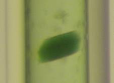
Figure 1. Crystals of Higher Plants Photosystem II.