Magnetron deposited TiO2 thin films -crystallization and temperature dependence of microstructure and phase composition
R. Kužel1, L.
Nichtová1, Z. Matěj1,
J.
Šícha2, J. Musil2
1Department of Condensed Matter Physics, Faculty of Mathematics and Physics, Charles University in Prague, Ke Karlovu 5, 121 16 Praha 2, Czech Republic
2Department of Physics, Faculty of Applied Sciences,
University of West Bohemia in Pilsen, Czech Republic
* Contact author; e-mail: kuzel@karlov.mff.cuni.cz
Keywords: powder diffraction, titanium dioxide, crystallization, nanocrystalline thin films
Titanium dioxide ( TiO2 ) films are nowadays widely used because of their interesting photocatalytic and self cleaning properties. Complex X-ray scattering studies were performed on sets of titanium dioxide thin films sputtered by dual dc magnetron [1]. Several sets of nanocrystalline and amorphous TiO2 thin films magnetron deposited on glass and silicon substrates have been studied. Phase analysis and X-ray line broadening were studied by X-ray powder diffraction in parallel beam optics; the residual stresses were measured with the aid of the Eulerian cradle and surface roughness determined by X-ray reflectivity measurement. Microstructure parameters were extracted from XRD measurements by individual peak profile fitting and also by whole powder pattern modelling [2] approach (MAUD [3], modified FOX[4]).
A set of amorphous films with different thickness was studied after annealing and also by in-situ measurements during the heating. It was found that the crystallization temperature started at about 250 °C for thicker films but it was higher for thinner films (< 200 nm) and reached about 350 °C. Thinner films were single phase (anatase) while thicker films above 1200 nm contained also a small amount of nanocrystalline rutile. The crystallite size of these samples immediately after crystallization was larger than 100 nm.
This is clearly different from as-deposited nanocrystalline films. By both thickness dependence of XRD patterns and depth profiling measurements it was found that rutile phase growths on the substrate and it is transformed to anatase with increasing distance from the substrate. This may be caused by temperature gradient during the deposition. Thinner films consist mainly of about 7 nm large rutile crystallites. With annealing temperature the crystallite size is continuously increasing (Figure 1a). By contrast, in thicker films the anatase crystallites of similar size (7 nm) dominate but they remain nearly unchanged with annealing up to 500 °C and then they are transformed to larger rutile crystallites after annealing at 800 °C (Figure 1b).
Simple uniaxial tensile stress and only a weak texture were found for the amorphous films after crystallization. Only for the thinnest films (~ 100 nm), the 101 texture (anatase) was found. In case of nanocrystalline films the stress was low but not uniaxial. This is related to significantly stronger and more complicated texture due to the dual magnetron geometry.
Reflectivity measurements revealed increasing surface roughness with both increasing thickness and annealing temperatures.
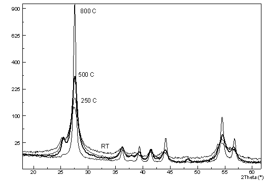
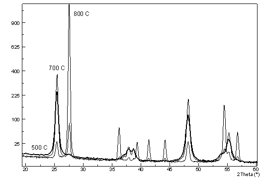
a) b)
Figure 1. Diffraction patterns of TiO2 films annealed at different
temperatures. Strongest peaks of anatase and rutile are 101 at 25° and 110 at
27°, respectively – a) thinner film (220 nm) with dominating rutile and
gradually increasing crystallite size with annealing (500 °C, thick line), b) thicker
film (935 nm) with dominating anatase after deposition and nearly unchanged crystallite
size up to 500 °C (thick line). Anatase is transformed into rutile after
annealing at 800 °C. Rutile peaks appeared already after annealing at 700 °C.
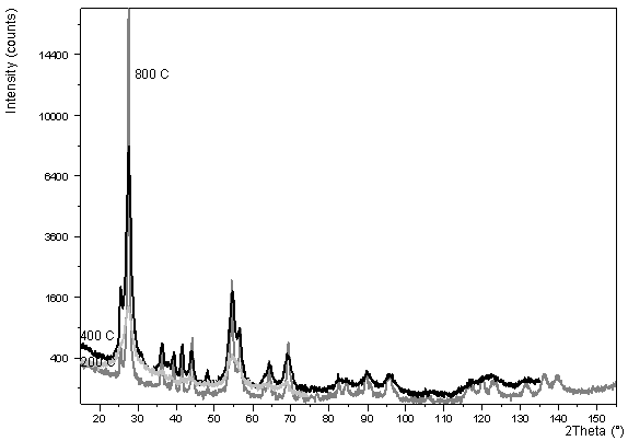
Figure 2. Diffraction pattern of TiO2 film with thickness 100 nm
annealed at different temperatures (200 °C, 400 °C, 800 °C).
1. J. Musil, D. Heřman, J. Šícha (2006). J. Vac. Sci. Technol. A24(3), 521-528.
2. P. Scardi, M. Leoni (2002). Acta Cryst. A 58, 190-200.
3. L. Lutterotti, www.ing.unitn.it/~maud.
The work is a part of the research programs MSM 0021620834 and MSM 4977751302 financed by the Ministry of Education of the Czech Republic and also supported by Grant Agency of the Czech Republic (106/06/0327).