Micro-diffraction with a mono-capillary: how to setup our experiment
P. Bezdička, E. Kotulanová
petrb@iic.cas.cz
The experimental setup of a micro-diffraction experiment has already been described elsewhere [1]. One of the most important aspects of the micro-diffraction experiment is the alignment of a sample.
The spot on the sample that is to be analyzed can be determined by means of an alignment microscope. This microscope in attached to a PreFIX interface and it is equipped with a cross-hair in the ocular. This setup permits one to adjust samples with a precision of about 50 mm. There is no way how to store the “in situ” information about the analyzed point or even about the precision of the system alignment.
Therefore we decided to modify this experimental setup using the alignment microscope. Together with IntracoMicro, Ltd. we constructed an optical interface for accommodation of a video camera in the position of the microscope eyepiece (fig. 1)
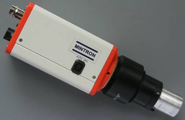
Figure 1: Mintron MTV-62W1P camera equipped with an optical interface
This interface is equipped with a similar cross-hair that is also aligned with the optical system of our X’PertPRO diffractometer. Therefore it could be used in the same way as an optical eyepiece. Either an analog or a digital video camera with a ½” sensor and C/CS lens can be used with this interface. The choice of a camera depends on what is the main purpose of the use of such attachment. If it is the preference of a good documentation of experiments, a digital camera may be in preference, as it permits the production of photographs with a better resolution and better reproduction of colors. If it is necessary to check the alignment of the system that needs to visualize the trace of the primary beam on the surface of a fluorescence disk, the analog camera, with its superior sensitivity, is absolutely necessary. The use of such a camera (with the sensitivity better than 0.01 Lux) can be very useful for a routine check of the system. An inferior resolution and color in accuracy are the drawbacks of that choice.
After consideration of all benefits and drawbacks of the use of either an analog or a digital camera, we decided to install for our system the analog “Mintron MTV-62W1P” camera (fig. 1) with the minimum sensitivity of 0.007 Lux.
The overview of the experimental setup installed on our X’PertPRO diffractometer is shown in the figure 2.
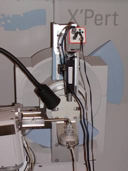
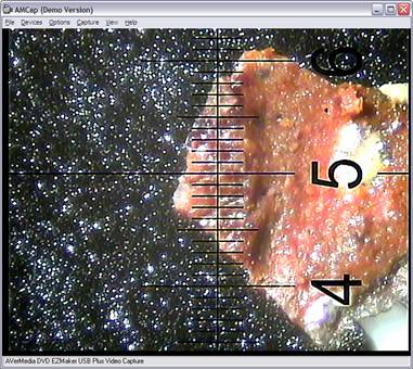
Figure 2: A video camera installed in the Figure 3: A typical photograph of the aligned sample
X’PertPRO diffractometer that is to be analyzed.
Figure 3 shows a typical sample of a fragment that has been placed on a Si zero background sample holder and set up for X-ray powder micro-diffraction with the analyzed point (cross-hair)
The X-ray micro-diffraction with a
conventional X-ray tube, focusing mono-capillary with a diameter of
References
1. V. Šímová, P. Bezdička, J. Hradilová, D. Hradil, T. Grygar, Powder Diffract., 20, (2005), 224.