Introduction
Synchrotron radiation topography is used for the investigation of many types of domains, phase coexistence, or field related defects in magnetic and ferroelectric single crystals. The origin of the contrast either resides in i) the variation of electrostrictive, magnetostrictive, or space charge related, distortions, or, ii) by taking advantage of the coherence of the beam, from a contrast mechanism associated with the variation of the structure factor phase between neighbouring domains.
Ferroelectric crystals: the example of KTP
We are concerned with ferroelectric 180░ domain walls, that behave as af twin boundary that separates regions of opposite polarity.
The aim of this study was to understand the structure of
domain walls in KTiOPO4 (KTP) crystals and to extract from coherent
X-ray Bragg diffraction imaging a very elusive information about atomic
arrangements at domain walls. We used for this a combined Bragg and Fresnel imaging technique that takes
advantage of the coherence of the synchrotron X-ray beam, and was succesfuly
used for LNO [1] and KTA [2] crystals. The domain walls were introduced by the
method of periodic poling. This means that the
ferroelectric polarization (and the crystal structure) is inverted in the
created neighbouring domains. X-ray diffraction by these, neighbouring ōupö and
ōdownö domains differ both through the amplitude and phase of the structure
factors. However, by far the largest contribution to the contrast between the
domains is produced by the difference in phase, which is principally structural
in origin. The value of this ōphase jumpö, Dj calculated
from the crystal structure depends very sensitively on how the crystal
structures of the domains are matched or linked across the domain wall. From
crystallographic principles, five possible domain‑matching schemes have
been suggested for KTP crystals. Each of these matching schemes introduces a
different phase shift in Bragg diffraction from inverted domains. Therefore, by
measuring the actual value of Dj and comparing it with values calculated from these models,
atomic‑level information about the domain wall can be inferred.
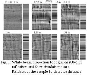
Fig. 1 shows reflection topographs of a 9 mm period KTP sample using the 004 reflection. The contrast simulations are presented on the right
side of the each experimental image. The
simulations were performed for Dj = ‑38.6░,
expected if the P(1) atom acts as a pivot for the twinning. Fig. 2 shows
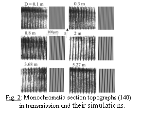
the set of monochromatic section topographs in transmission of a periodically
poled KTP sample with 24.7 mm period
as a function of the sample-to-detector distance, using the 140 reflection. The
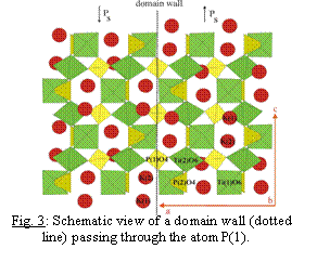
simulated images correspond to Dj = 180░. The model of twinning resulting from our investigation is
shown in Fig. 3. The translation ½ (a+b) relates equivalent atoms in adjacent domains in addition
to the shift in the c-direction [3].
Magnetic crystals: a-Fe2O3 and MnP
We will give two examples of magnetic phase transitions, where real time X-ray topography provides physical information that is not available otherwise.
Spin-reorientation
Morin transition in hematite Hematite (a-Fe2O3)
is a weak ferromagnet at room temperature. It undergoes a spin-reorientation
transition (the Morin transition, TM≈260K) towards a low
temperature antiferromagnetic state. Previous investigations indirectly suggested
that the boundaries that form during this transition are nearly parallel to the
(111) plane. The present study was motivated by the possibility of using high
energy X-ray section diffraction
imaging, which allows the direct visualization of the boundaries along the
depth of a thick sample. The Morin
transition was therefore visualized by observing phase boundary movements under
the influence of temperature and of a magnetic field, on white beam section
topographs, with the FreLoN camera as detector. The sample was a high quality
(111) platelet shaped crystal, 1.1 mm thick. Figure 4 shows the phase boundaries
movement within the topographic images (corresponding to virtual slices of the
sample), while remaining nearly parallel to (111). These images were recorded
at a fixed temperature, within the antiferromagnetic phase (258 K) whereas
increasing the magnetic field (60 mT ,Ā
180 mT and 235 mTĀ in fig. 4) that
favours the weak ferromagnetic phase. The nucleation of the weak ferromagnetic phase and pinning of interphase
boundaries on defects located in the bulk of the crystal were observed. The
observed behaviour patterns were explained in terms of the elastic and
magnetostatic energies involved [4].
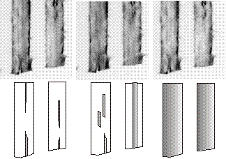
Fig; 4: Evolution of wo
section topographs recorded during the Morin transition as a function of the
magnetic field. The width of a given section corresponds to 0.35 mm. The
schematic representation of the section topographs shows the progression of the weak ferromagnetic phase,
(shaded regions).
Fan
to ferromagnetic transition in MnP
MnP can be produced as highly perfect single
crystals. It exhibits a complex magnetic phase diagram, which easily allows
alterations in the ratio between the various energy terms relevant for the
phase coexistence. The ferromagnetic - fan coexistence was found [5] to display
a thick, interface between the two magnetic phases (figure 5). Complementary
investigations were performed on a (001) MnP crystal, under a magnetic field
applied along the b direction, at T ≈ 48 K, in order to
understand this unusual phase boundary.
It was observed that the ferromagnetic-fan interface includes bulk transition regions, elongated along a, and thick enough along the b direction (in the 10-4 m range) to produce a substantial contribution to diffraction. The Bragg condition changes continuously across these regions. This configuration, which involves magnetic charge distribution, is in sharp distinction with the usual two-dimensional character of magnetic walls and phase boundaries. A model of the thick interface comprising a set of intermediate magnetic states that only occur during the ferromagnetic-fan phase coexistence, was proposed to explain the observations [6]. The very unusual fact is that, if this model applies, this thick interface is not expected to imply a substantial increase in elastic energy, because locally each magnetic state exhibits its spontaneous distortion, ideally leading to no long-range stress.
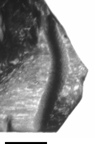
Fig; 5: Monochromatic
topographĀ recorded during the ferro to
fan transition, which shows the thick phase boundary (a is vertical and b
horizontal; scale bar: 1 mm).
- Rejmßnkovß-Pernot P, Cloetens P, Baruchel
J, Guigay J-P and Moretti P 1998 Phys. Rev. Lett. 81 3435.
- Rejmßnkovß-Pernot P, Thomas P A,
Cloetens P, Lorut F, Baruchel J, Hu Z W, Urenski P and Rosenman G 2000 J.
Appl. Cryst. 33 1149.
- Pernot-RejmßnkovßĀ P, Thomas P A, Cloetens P, Lyford T and
Baruchel J 2003 J.
Phys.:Condens. Matter 15 1613.
4.
Schetinkin S.A., Wheeler E., Kvardakov V.V., Schlenker M.and Baruchel J.
2003 J. Phys. D:
Appl. Phys 36,Ā A118-A121
5.
Medrano C., Pernot E., Espeso J., Boller E., Lorut F.and Baruchel J. 2001
Journal of Magnetism and Magnetic Materials 226-230,Ā 623-625
6.
Baruchel J., Medrano C.and Schlenker M 2005 J.
Phys. D: Appl.
Phys 38, A67-A72