CHARACTERISTICS OF DUERR
IMAGING PLATE OPG
Z. Pala1,
N. Ganev1, K. Kola°Ēk1
1Department of Solid State
Engineering, Faculty of Nuclear Sciences and Physical Engineering, Czech
Technical University in Prague, Trojanova 13, 120 00
Prague 2, Czech Republic
Keywords
Imaging
plate, density of blackening, linearity, photographic film, backscattering
Introduction
The goal of
this study is to provide a relation between density of blackening and exposure
for Duerr Imaging Plate OPG, which is used as a
position sensitive detector in XRD laboratory of Department of Solid State
Engineering. Comparison between photographic film and imaging plate from the
point of view of linearity was performed. Backscattering Debye
¢ Scherrer experiment with Ag standard was carried
out in order to gain diffraction pattern on imaging plate. Dependences of background,
absolute and relative peak height versus exposure time for two wavelengths of
X-ray radiation were evaluated.
Density
of blackening
Absorption
of photon in the sensitive layer of silver bromide leads to formation of
photographic latent image. The unexposed crystallites of silver bromide are
removed in the fixing bath. Density of blackening D can be expressed as
D = log I0/I, ĀĀĀĀĀĀĀĀĀĀĀĀĀĀĀĀĀĀĀĀĀĀĀĀĀĀĀ (1.1)
where I0
is the intensity of incident light and I is the intensity of light that passed
through a developed and fixed photographic film. Density D may be
expressed as a function of exposure time t and intensity of incident
X-ray beam IX. Hence the characteristics of film is given by D
= f (IX*t), the maximum value of D where D is
linear function of IX*t varies from 0.5 to 2.5 [1].
Sensitive
layer in imaging plate comprises of luminofore barium
chromo-bromide, which is excited by incident photon into a semi-stable state.
By an illumination with He-Ne laser the process of photostimulated luminiscence is triggered
and the image in form of 16-bit grayscale pattern is
released. Now I0 in eq. (1.1) is the
maximum value on grayscale 216 = 65536 and
I is the information in the chosen pixel. It can be
still assumed that D is function of exposure time t and intensity
of incident X-ray beam IX.
Experiment
The
backscattering diffraction experiment was done using CuKa and CrKa radiation, the exposure times
varied from 1 to 20 minutes for copper anode and from 1 to 55 minutes for
chrome anode. The plate holder was rotated at 1 rpm in order to avoid effects
of coarse grain of the standard Ag. The incident beam impinged the sample in
direction normal to its surface. The 16-bit diffraction pattern on the imaging
plate was obtained by scanning on VistaScan by Duerr. Lucia 5.10 image analysis system was used to gain
intensity profile, which was transformed into density profile employing eq. (1.1).
Evaluation
of profiles
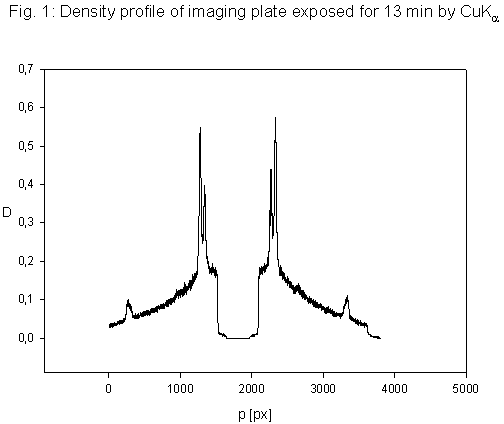
In Fig. 1
density profile is depicted, dependences of following parameters on exposure
time were investigated:Ā absolute and
relative diffraction peak height, integration intensity, FWHM and level of
background. The peaks were approximated by Gaussian function D = a + b*exp(-0,5((p-c)/d)2),where p is
position in pixels, and background by two linear functions D = K1p+Q1,
D = K2p+Q2. Value Dlin
as a maximum density, where linear relation between D and t is
observed, was figured out for both wavelengths.
Ā
Conclusions
Following
statements can be derived from computed characteristics of imaging plate:
(i)
Value Dlin
= 0.3 was established. That corresponds to the intensity range
(34000, I0) in 16-bit image.
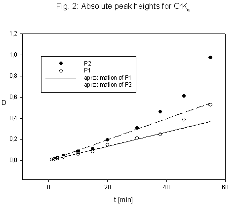
(ii)
Photographic films exhibit effect of solarization for D > Dlin,
when the level of blackening declines by big exposures. Whereas imaging plate
display higher values of density for D > Dlin
than would correspond to linear evolution as can be seen in Fig. 2.
(iii)
If the absolute peak height is less than Dlin, the plots Iint (t), b (t) are linear. ForĀ D >
Dlin deviations from linearity
occur.
(iv)
No obvious relation between FWHM and exposure was
found.
References
1.ĀĀĀĀĀĀĀĀ Kraus I., Ganev N.: Technickķ aplikace difrakĶnĒ anal²zy. Praha 2004. VydavatelstvĒ ╚VUT.
2.ĀĀĀĀĀĀĀĀ Giacovazzo C., et al.: Fundamentals of Crystallography.
Second edition. Oxford University Press 2002.