SAXS AND QELS STUDY OF Au AND UHMWPE PARTICLES
1M. Šlouf, 1J. Pleštil, 1H. Synková, 2J. Kumstátová, 2S. Eklová
1 Institute of Macromolecular Chemistry, Academy of Science of the Czech Republic, Heyrovského náměstí 2, 16206 Praha, Czech Republic; slouf@imc.cas.cz
2Department of Biochemistry, Faculty of Science, Charles University, Hlavova 8, 12840 Praha 2, Czech Republic
Introduction
Total replacement of human hip joints, or total hip replacements (THR), usually have two main components: the first is metallic and the second is made of UHMWPE (ultrahigh molecular weight polyethylene). This polymer is regarded as the best possible material for the application due to its excellent friction and satisfactory mechanical properties [1]. Nevertheless, articulation of the metallic and polymeric component in THR leads to wear, i.e. formation of tiny UHMWPE particles, whose size varies around 1 mm. The particles are released to the surroundings of THR, where they induce inflammatory reactions leading to osteolysis. The whole process is believed to be the main cause of THR failures [2].
One of the tasks in our project is to determine the number of UHMWPE wear particles in zones around THR and correlate the number of particles with the extent of tissue and bone damage in these zones [3]. As the wear particles are almost invisible even under light microscope, their quantification is not easy. Isolation of the wear particles and determination of their weight is possible but it is quite imprecise due to their negligible size [4]. That is why we tested several other techniques, which were not based on particles weighing. In this study we present a technique relying on quasi-elastic light scattering (QELS) of the particles in solution. QELS signal is quite strong even if the studied suspension contains unweighable amount of particles. However, standard QELS experiments do not yield the numbers of the particles, only their intensity distribution on relative scale. Size range of the method is approximately from 1 nm to 6 mm, and the wear particles have dimensions ³ 0.1 mm. Therefore we need to add to the suspension of UHMWPE particles a known amount of calibration particles smaller than 100 nm, for example gold nanoparticles of suitable size [5]. Theoretically, QELS scattering of a solution containing UHMWPE wear particles and Au calibration particles should give two-peak distribution curve. The unknown amount of the UHMWPE particles could be calculated from the known amount of the Au particles and relative intensity of the two peaks.
The principle of the method, which we called QELSc (i.e. QELS with calibration particles), and which is briefly described in the previous paragraph, is quite simple. From the theoretical point of view, QELSc should work perfectly. From the practical point of view the method has several limitations, the most important of which are summarized below.
Experimental
Preparation of Au nanoparticles. The colloidal solutions of Au nanoparticles with pre-calculated size were prepared by controlled reduction of H[AuCl4]. The principle of the preparation was described as early as the last century [6]; our slight modification of this technique, allowing pre-calculation of the prepared particles, appeared recently [5,7].
Preparation of UHMWPE wear particles. UHMWPE wear particles for testing were prepared in vitro. UHMWPE powder was put into liquid nitrogen and pulverized so that very small particles were obtained. The powder was suspended in propan-2-ol (= isopropyl alcohol, iPrOH) and suspensions of the particles with sizes less than 0.1, 1.0 and 5.0 mm were prepared by filteration through polycarbonate microfilters (Whatman).
Transmission electron microscopy (TEM) of Au nanoparticles was carried out with microscope JEM 200CX (Jeol). All TEM microphotographs were taken at acceleration voltage 100 kV and recorded with digital camera (MegaView).
Quasi-elastic light scattering (QELS) was measured with Zetasizer Nano ZS (Malvern Instruments Ltd.) in the size mode. Intensity distribution curves were calculated with a general model, without any restrictions imposed on the measured data. [8]
Small-angle X-ray scattering curves (SAXS) were obtained with an upgraded Kratky camera equipped with a position-sensitive detector. The number distributions of the particle radii were calculated from smeared scattering data using Glatter’s desmearing program ITP [9].
Results
and discussion
The main goal of this study was the verification of QELSc method. In the first step, a set of colloidal solutions of Au nanoparticles with different sizes were prepared. The theoretical sizes of the nanoparticles were pre-calculated before the experiment [5,7] and the real sizes of Au nanoparticles were determined by image analysis of TEM microphotographs (Fig. 1).
|
|
|
|
|
(a) |
(b) |
(c) |
|
|
|
|
|
(d) |
(e) |
(f) |
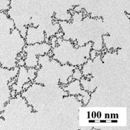
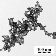
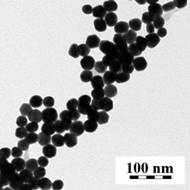
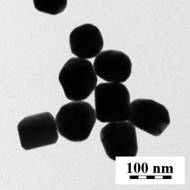
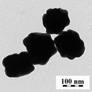
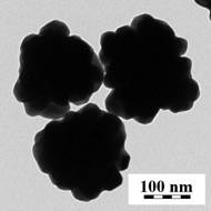
Fig. 1. TEM micrographs of Au nanoparticles: (a) colloid Au1, (b) Au2, (c) Au3, (d) Au4, (e) Au5 and (f) Au6. The average sizes of the colloids Au1, Au2, Au3, Au4, Au5 and Au6 are ca. 4, 10, 30, 100, 150 and 200 nm, respectively.
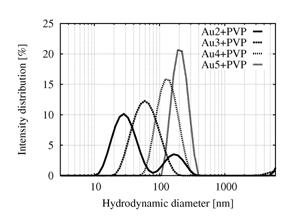
In the second step, the nanoparticles were stabilized with PVP (polyvinylpyrrolidone) and their QELS curves were measured (Fig. 2). Colloids Au1 and Au6 are not shown in Fig. 2 because their QELS curves were unusable. Colloid Au1 contained the highest concentration of nanoparticles and so its intensity distribution was distorted due to formation of agglomerates. The impact of agglomeration on the QELS intensity distribution curve is very strong because each point of the curve is proportional to Nd×d6, where d is the particle diameter and Nd is the number of particles with diameter d. Consequently, the large particles and/or agglomerates dominate the intensity distribution even if their concentration is quite low. The agglomerates are also observed in the QELS curve of Au2 in the form of the second, lower peak. The intensity distribution of colloid Au3 did not exhibit any agglomeration, which accords with the fact that the concentration of nanoparticles decreases with their increasing size; this results from our preparation procedure [5,7]. Colloid Au6 contains the largest and heaviest nanoparticles, whose fast sedimentation was visible even with the naked eye: within several tens of minutes a layer of Au6 nanoparticles appeared at the bottom of the flask. Generally speaking, Au6 had not exhibited colloidal behavior any longer and this destroyed the QELS curve.
|
|
|
|
Fig. 2. QELS intensity distributions of colloids Au2, Au3, Au4 and Au5. |
Fig. 3. SAXS number
distributions of colloids Au1, Au2,
Au3 and Au4. |
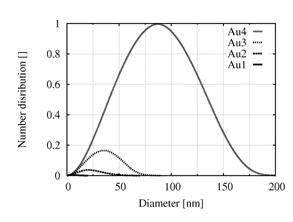
In the third step, SAXS and QELS curves of colloids Au1, Au2, Au3 and Au4 were compared to get insight into particles behavior in solution. SAXS curves of Au5 and Au6 colloids could not be measured because their nanoparticles exceeded the SAXS resolution (1 - 100 nm). A comparison of intensity distribution curves from QELS with standard number distribution curves from SAXS proved that the unusual shape of Au1 and Au2 intensity distribution curves could be accounted for agglomeration, which is overestimated in QELS.
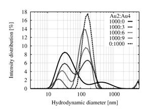
In the fourth step, various pairs of colloids with different sizes were mixed. The best results were obtained with a mixture Au2:Au4; relative intensity of the peaks changed in agreement with changing Au2:Au4 ratio (Fig. 4). The concentration of Au2 particles had to be much higher because of their smaller dimensions resulting in their much lower scattering power. The concentration of Au4 particles had to be kept within quite a narrow range so that both peaks could be observed simultaneously. Other combinations were not so successful, which illustrates the limitations of QELSc technique. Colloids Au1 and Au6 were excluded at the very beginning because their QELS curves were unusable as described above. Combination of Au3:Au4 did not give reasonable results because the size of the colloids is too close and, as a result, the QELS curve of the mixture exhibited just one peak. The same was true for a Au3:Au5 mixture. Combination Au2:Au5 could have been used as well but the disadvantage in this case was too large difference in particle sizes: as the QELS signal is proportional to six power of particle diameter, the abundance of colloid Au2 in comparison with Au5 would have had to be too high.
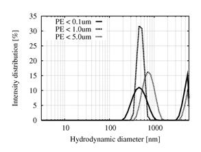
In the fifth step, in vitro UHMWPE particles of different sizes were prepared as described in the experimental section and the QELS curves of their suspensions in iPrOH were measured (Fig. 5). The intensity distribution curves proved that PE particles give sufficient QELS signal. No agglomerates were observed. Surprisingly, the intensity distributions of all three suspensions, each of which contained different particles, were more-or-less the same. This may have been caused by inappropriate preparation of the particles and/or the absence of their colloidal behavior due to unsuitable solvent. The wrong preparation procedure may have lead to the particles of the same size, which will be checked by scanning electron microscopy of the isolated particles on polycarbonate membrane. The unsuitable solvent could also influence the results, because r(iPrOH) = 0.78 g/cm3 is significantly lower than r(UHMWPE) = 0.94 g/cm3. As the UHMWPE particles were quite large, their sedimentation could have destroyed the QELS signal. This will be checked by QELS measurement of UHMWPE particles in a mixture of iPrOH and H2O with suitable density.
|
|
|
|
Fig. 4. QELS intensity distributions of the mixture of colloids Au2:Au4. |
Fig. 5. QELS intensity distribution curve of the in vitro UHMWPE wear particles. |
Conclusion
Determination of UHMWPE wear particles is not an easy task. The weighing of the particles is imprecise due to their tiny dimensions. That is why the authors of this work tried to develop a new technique, called QELSc, which is based on quasi-elastic light scattering. The results indicate that the principle of the method is correct although there are serious limitations concerning particle sizes, concentration ranges and precision. The comparison of SAXS and QELS measurements also confirms that the agglomeration of the particles is a crucial problem of QELSc. Experiments with Au nanoparticles proved that QELSc can be employed in estimating the number of unweighable amounts of particles. The first experiments with UHMWPE wear particles yielded QELS intensity distribution curves without agglomerates, but they seem to be insensitive to particle sizes, which will be a subject of further study.
Acknowledgement. This work was supported by the Grant Agency of the
Czech Republic (grants GACR 106/04/1118 and GACR 203/04/0688) and by the
Academy of Sciences of the Czech Republic (project AVOZ4050913).
References
1. S. M. Kurtz: UHMWPE handbook. San Diego, USA, 2004. Elsevier.
2. A. Bellare, A. Bistolfi, K. Simis & L. Pruitt, Proceedings of UHMWPE for arthroplasty. Torino, Italy, 19.5.2003, pp.33-51.
3. M. Šlouf, I. Šloufová, G. Entlicher, Z. Horák, M. Krejčík, P. Štěpánek, T. Radonský, D. Pokorný & A. Sosna, J. Mater. Sci.- Mater. Med. 15 (2004) 1267-1278.
4. S. Eklová, G. Entlicher, H. Synková, M. Šlouf & A. Sosna: Reliable Technique for Isolation of Polyethylene Wear Debris, in preparation.
5. M. Šlouf, R. Kužel & Z. Matěj: Materials Structure, 11 (2004) 166-168.
6. J. Turkevich, P. C. Stevenson & J. Hillier, Disc. Faraday Soc., 11 (1951) 55-75.
7. H. Vlková, M. Šlouf, Č. Koňák, J. Pleštil, J. Hromádková & H. Synková: Proceedings of Mikroskopie 2005, Nové Město, Česká republika, p. 44.
8. Zetasizer Nano Series User Manual. Malvern Instruments Ltd. 2003, 2004.
9. O. Glatter in: Neutrons, X-rays and Light: Scattering Methods applied to Soft Condensed Matter (P. Lindner & Th. Zemb, editors), Amsterdam, Netherlands, 2002. Elsevier. pp. 103-124.