Comparative
study of porosity in 3Y-TZP superplastic ceramics by USANS and SEM image
analysis
V. Ryukhtin1,2, J. Šaroun1,
S. Harjo3, Y. Motohashi3, M. Baron4,5, R.
Loidl4,5
1-
Nuclear Physics Institute, 25068 Řež near Prague, Czech Republic.
2-
Charles University, Faculty of Mathematics and Physics, Ke Karlovu 3, 121 16
Praha 2, Czech Republic.
3-
Ibaraki University, Faculty of Engineering, The Research Center for
Superplasticity, Hitachi, Ibaraki, 316-8511 Japan.
4-
Institute Laue-Langevin, BP 156, F-38042 Grenoble Cedex 9, France.
5-
Atominstitut der Österreichischen Universitäten, A-1020 Wien, Austria.
Evolution
of cavities in superplastic ceramics is a significant indicator of mechanism of
the superplastic deformation process. Moreover the cavities have large
influences on mechanical and functional properties [1]. The cavities and pores
in fine-grained 3Y-TZP have sizes typically between 0.1 mm and 1 mm and can therefore be
studied by both scanning electronic microscopy (SEM) image analysis and ultra small-angle
neutron scattering (USANS). However, quantitative comparison of results
obtained by the two techniques is complicated by surface artefacts, uncertainty
in pore identification, assumptions of the model used to fit the USANS data and
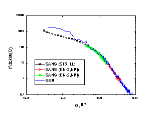
other factors. We show that scattering functions measured at double-crystal
USANS instruments can be calculated directly from sufficiently large SEM images
of the sample sections parallel to the scattering plane. The results of both
techniques can therefore be compared directly. The porosity in 3Y-TZP ceramics
samples deformed from 0% to 200% were measured in broad range of Q (2·10-5
– 8·10-3 Å-1) using Bonse-Hart SANS (S-18 at
ILL, Grenoble) and double crystal (DC) SANS (DN-2 at NPI, Řež near Prague)
instruments. The measured scattering curves were compared with the scattering
curves, calculated directly from the SEM images. Remarkable agreement of both
methods was observed (Fig. 1), except of small Q-range, where the contribution
of very large pores prevails. For such big pores, numerical and statistical
errors of calculation of the scattering function from SEM images become large
[2].
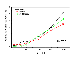
USANS
experiments enabled to measure alternatively the bulk porosity with the results
consistent to those of other techniques (SEM, Archimedes) (Fig. 2). The
presented correlation between measured data and calculated from SEM images
indicates, that the SEM images represent well the bulk microstructure of the
material and the pore identification procedure doesn't introduce significant bias
to the SEM results for pores with radii <1 mm. However, at strains near
200%, large cavities (R~5mm) have been observed on some SEM images. Comparison with SANS
indicates, that these cavities are either surface artefacts or are not
distributed in the whole bulk investigated by SANS.
Fig. 1. Comparison of measured data with scattering function calculated
from SEM images. Fig. 2. Comparison between the volume fraction of
cavities measured by SANS, those by image processing and those by
Archimedes method.
References
[1] Y. Motohashi, N. Sugeno, S. Koyama and T. Sakuma, Mater. Sci. Forum,
243-245, 1997, pp. 399-404.
[2] V. Ryukhtin, J. Šaroun. Direct comparison of SANS data with SEM
image analysis. Proc. ICNS-2001. J. Appl. Phys. A. (in print).