Cryo-EM structure of the large photosynthetic complex from two species of Gemmatimonadota
Alastair T. Gardiner1, David Bina2, 3, Pu Qian4, Zdenko Gardian2,5, David Kaftan1,2 & Michal Koblížek1*
1Institute of
Microbiology of the Czech Academy of Sciences, 379 81 Třeboň, Czech Republic.
2Faculty of Science, University of South Bohemia, 370 05 České
Budějovice, Czech Republic.
3Biology Centre, Czech Academy of Sciences, Institute of Plant
Molecular Biology, Branišovská 1760, 370 05 České Budějovice, Czech Republic.
4Materials and Structure Analysis, Thermofisher
Scientific, Achtseweg Noord 5, 5651 GG Eindhoven, Netherlands.
5Biology Centre,
Czech Academy of Sciences, Institute of Parasitology, Branišovská 1760, 370 05
České Budějovice, Czech Republic.
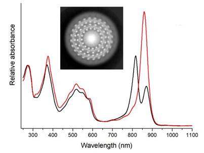
Members of the bacterial phylum Gemmatimonadota (previously called Gemmatimonadetes) inhabit a wide and diverse range of habitats including soils, aquatic environments i.e., marine, fresh and waste water as well as biofilms and sediments. In spite of the prevalence in Nature of this phylum only six cultured species have been described so far. Two of these species, Gemmatimonas (Gem.) phototrophica and Gem. groenlandica are photoheterotrophic and contain anoxygenic photosynthetic complexes. The photosynthetic pigment-protein complex in Gem. phototrophica was found to contain two concentric antenna rings around the central reaction centre (RC), (a projection image is inset in the Figure), and we obtained a high resolution, cryo-EM structure followed revealed many novel features [1].
Due to the double-ringed antenna in this complex it was termed RC-dLH (d = double Light-Harvesting (rings)). Gem. groenlandica was the second phototrophic Gemmatimonadota strain to be characterised [2]. Intriguingly, the absorption spectrum of Gem. groenlandica RC-dLH complex (red line) is rather different to that of Gem. phototrophica (black line) in the near infra-red (NIR), see Figure. We are currently in the process of final refinement for the RC-dLH complex from Gem. groenlandica, therefore, this presentation will compare and contrast the two structures and provide possible functional and physiological reasons for these differences.
[1] P. Qian, A.T. Gardiner, I. Šímová, K. Naydenova, T.I. Croll, P.J. Jackson, Nupur, M. Kloz, P. Čubáková, M. Kuzma, Y. Zeng, P. Castro-Hartmann, B.v. Knippenberg, K.N. Goldie, D. Kaftan, P. Hrouzek, J. Hájek, J. Agirre, C.A. Siebert, D. Bína, K. Sader, H. Stahlberg, R. Sobotka, C.J. Russo, T. Polívka, C.N. Hunter, M. Koblížek, 2.4 Å structure of the double-ring Gemmatimonas phototrophica photosystem, Science Advances, 8 (2022) eabk3139.
[2] Y. Zeng, Nupur, N. Wu, A.M. Madsen, X. Chen, A.T. Gardiner, M. Koblížek, Gemmatimonas groenlandica sp. nov. Is an Aerobic Anoxygenic Phototroph in the Phylum Gemmatimonadetes, Frontiers in Microbiology, 11 (2021).