Atomic force microscopy in structural biology
J. Přibyl1, Š. Klimovič1, R. Obořilová1, D. Kabanov1, J. Mlčoušková2
1CF NanoBiotechnology, CEITEC MU, Masaryk University, Brno, Czech Republic
2LORD Group, Faculty of Science, Masaryk University, Brno, Czech Republic
jan.pribyl@ceitec.muni.cz
Atomic force microscopy (AFM) can generate images within ranges of resolution that are of particular interest in biology [1]. Although atomic resolution may not be possible with biological samples, a great deal of information can still be obtained from images that provide structures at a slightly lower level of resolution.
Our laboratory is performing research in the field of imaging of biomolecules [2], mapping the elastic properties of cells and their clusters [3], and characterization of contractile properties of cardiomyocytes and their clusters [4].
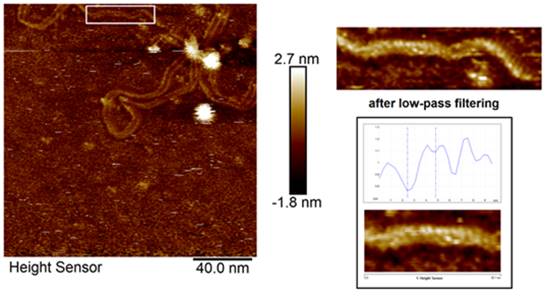
Figure 1. DNA structure studied by the AFM.
The laboratory's flagship is the large AFM microscope JPK NanoWizard 4XP installed on a Leica DMi8 optical microscope with a fluorescence module. Both microscopes can operate simultaneously in the so-called directoverlay mode, thus combining AFM and optical microscopy abilities. Moreover, this microscope is not only an imaging tool, however helps to map elastic properties of various samples with nanometer resolution. One of the main advantages is the ability to work in semi-physiological conditions.
Combining the AFM with microfluidic, so-called FluidFM enables the possibility to aspirate and/or deliver extremely low volumes. This feature can be used when injecting or removing small volumes from individual cells. Using stiffer cantilevers, the system can investigate cell adhesion on new types of implant materials.
Keeping on the cutting-edge current AFM technology, the new generation of the MultiMode AFM microscope, version 8HR, was built for imaging with the maximum resolution that current commercial setups allow. This AFM setup will help the structural biologist image the biomolecules (DNA, proteins, molecular complexes) on a single molecular level.
Moreover, the multielectrode array (MEA) can be simultaneously connected with an AFM microscope, thus studying mechanoelectrical feedback of cardiac cells, tightly connected with some heart pathologies, such as catecholaminergic polymorphic ventricular tachycardia (CPVT).
1. Czajkowsky, D. M., Iwamoto, H., & Shao, Z.: J. Electron Microscopy 49(3) (2000) 395–406.
1. Horňáková, V., Přibyl, J. & Skládal, P: Monatsh Chem 147 (2016) 865–871.
2. Raudenska M., Kratochvilova M., et al.: Scientific Reports 9 (2019) 1660, 1-11.
3. Caluori G, Pribyl J, et al.: Biosens Bioelectron. 124-125 (2019) 129-135.
CIISB, Instruct-CZ Centre of Instruct-ERIC EU consortium, funded by MEYS CR infrastructure project LM2018127 and European Regional Development Fund-Project "UP CIISB "(No. CZ.02.1.01/0.0/0.0/18_046/0015974), is gratefully acknowledged for the financial support of the measurements at the CF Nanobiotechnology.