Study of Ni2FeGa microwire by conventional X-ray diffraction
Petr Cejpek, Limpat Nulandaya, Ladislav Straka
Institute of Physics, Czech Academy of Sciences, Na Slovance 1999/2, 182 00 Praha 8, Czech Republic
Ni2FeGa is a magnetic shape memory alloy with potential applications in microdevices such as actuators and micropumps. The preparation of bulk single crystals of this material is challenging; however, the fabrication of microwires containing highly oriented grains appears promising and economically feasible, as large quantities of such wires can be produced [1]. Still, the characterization of these microwires by conventional X-ray diffraction remains difficult due to their small diameter (typically 10–50 μm), which results in a limited diffracting volume. An additional challenge is the manipulation and mounting of the sample.
The Ni2FeGa microwires were placed and stretched inside own 3D-printed sample holder (Fig. 1) and initial measurements were performed using a SmartLab diffractometer with a 2D pole figure method and a Hypix3000 area detector positioned as close to the sample as possible. This setup significantly reduces the acquisition time for a single pole figure. However, individual reconstruction of pole figures is required to avoid the integration of a large background contribution and diffraction peaks originating from the sample holder.
Several conclusions can be drawn from the pole figures: 1) the presence of both cubic austenite and tetragonal martensite phases can be identified; 2) Ni2FeGa exhibits a strong preferential orientation with the ⟨110⟩ direction aligned along the wire axis; and 3) the presence of single or multiple martensitic variants is clearly observable.
These initial results open the way for future in situ experiments, such as in situ Joule heating by passing an electric current through the microwire, or in situ mechanical straining using a dedicated sample holder with a geared mechanism.
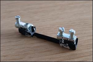
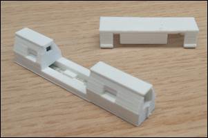
Figure 1. 3D-printed microwire holder. a) The first prototype, b) the second prototype – the metallic parts are placed upside down inside the plastic body to hide the screws.
[1] Frolova, L., et al. Reversible structural transition in monocrystalline Ni2FeGa microwires for shape-memory applications, Materials Science and Engineering B 263 (2021) 114891, https://doi.org/10.1016/j.mseb.2020.114891.