Magnetic morphology of multishell nanoparticles
Š. Hricov1, O. Kaman2, I. Dirba3, N. J. Steinke4, D. Zákutná4,5
1Department of Condensed Matter Physics, Faculty of Mathematics and Physics, Charles University, Ke Karlovu 5, 121 16 Prague 2, Czechia
2Institute of Physics of the Czech Academy of Sciences, Cukrovarnická 10, 162 00 Prague 6, Czechia
3 Technical University of Darmstadt, Karolinenplatz 5, 64289 Darmstadt, Germany
4Institut Laue-Langevin, 71 Avenue des Martyrs CS 20156, 38042 Grenoble, France
5Department of Inorganic Chemistry, Faculty of Science, Charles University, Hlavova 8, 128 00 Prague 2, Czechia
Magnetic nanoparticles (MNPs) are of high research interest due to their unique physical properties, which lay the foundation for various applications, ranging from biomedical diagnostics and therapeutic interventions to high-density data storage systems and environmental remediation processes. Broadly known and well-studied materials of this class are iron oxides MNPs. Among other research interests, they have been heavily exploited for their heating abilities via magnetic fluid hyperthermia. This process is a cornerstone for innovative cancer treatment therapies, which aim to localise tumour elimination. However, we propose a novel candidate material, the ε-Fe3N. It possesses unprecedented magnetic properties, essentially surpassing the well-established iron oxide MNPs [1], having larger saturation magnetization, leading to better heating performance in hyperthermia. As a result, the required therapeutic temperatures for tumour ablation can be achieved with a less concentrated MNP dispersion, thereby reducing the dose needed. Nevertheless, due to the nano-sized crystals, the ε-Fe3N is air sensitive, which results in massive oxidation. Thus, a robust surface protection must be realised. While considering the potential biomedical applications, we propose a silica encapsulation procedure to hinder ε-Fe3N oxidation and to establish biocompatibility, together with possibility to form aqueous dispersions. To successfully grow a silica layer, we have chosen a route of ε-Fe3N surface passivation, which we present in this contribution, together with insight into magnetic behaviour of complex core@shell MNPs. The bright-field transmission electron microscope micrographs (Figure 1: A) and small-angle X-ray scattering (Figure 1: B) show well-defined core@shell MNP morphology of the passivated nanoparticles with a mean particle diameter of 17.2(2) nm. Nevertheless, the macroscopic magnetization measurements revealed unexpected behaviour leading to a decrease in saturation magnetization and the presence of exchange bias at 5 K. To further explore, the complex magnetic nature of this material was disentangled by probing magnetic scattering fluctuations using the magnetic small-angle neutron scattering with incident beam polarization at the D33 instrument at ILL [2]. Finally, we will disentangle the magnetic morphology contributions from the magnetic core and shell part of passivated ε-Fe3N MNPs and discuss the resulting magnetic response of the presented MNPs in detail.
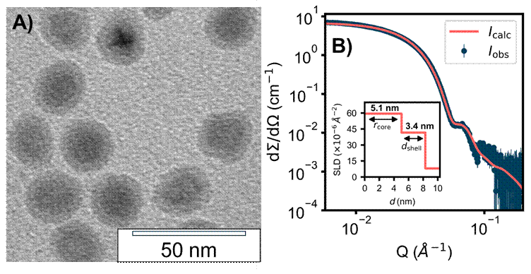
Figure 1: A TEM micrograph of passivated ε-Fe3N MNP, B SAXS curve recorded on dispersion of passivated ε-Fe3N MNP.
1. I. Dirba et al., J. Phys. D: Appl. Phys, 56, 025001 (2023). doi: 10.1088/1361-6463/aca0a9
2. Š. Hricov et al., Unmasking the Complex Core-Multishell Morphology of Magnetic Nanoparticles. Institut Laue-Langevin, proposal No. DIR-297 (2023). doi: 10.5291/ILL-DATA.DIR-297
This work was supported by the Czech Science Foundation (22-10035K) and the AMULET project, co-funded by MŠMT and the EU (CZ. 02.01.01/00/22_008/0004558). We also acknowledge the Institut Laue-Langevin for beamtime and financial support.