Synthesis and ab initio structure
determination from
powder diffraction data of K2ZrSi3O9.2H2O
A. Ferreira1, Z. Lin2,
M. R. Soares3, J. Rocha2
1 ESTGA, University of Aveiro, Apartado 473, 3754-909 Águeda, Portugal
2 Department of Chemistry, University of Aveiro, 3810-193 Aveiro, Portugal
3 Laboratório Central de Análises, Universidade de Aveiro, 3810-193 Aveiro, Portugal
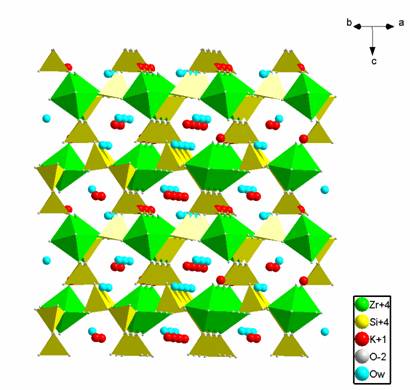
The crystal structure of a new potassium zirconosilicate K2ZrSi3O9.2H2O has been determined ab initio from powder X-ray diffraction data. The unit cell is orthorhombic, space group C2221 (no. 20), Z=4 with cell dimensions a = 8.1051(3), b = 10.6842(5), c = 12.0300(5) Å, V = 1041.76(7) Å3. The structure is made up of ZrO6 octahedra (MO6) and SiO4 tetrahedra (TO4) by sharing corners. The SiO4 tetrahedra form a helix chain, periodically repeating every six tetrahedral (step of 12.03 Å), along [001] interconnected by ZrO6 octahedra, forming a three-dimensional MT framework structure with channels delimited by seven and eight member rings. The potassium ions and water molecules are locate in this channels. The water molecules are reversibly lost on heating that is typical of zeolites and molecular sieves.
Experimental
Section
Synthesis. This phase was synthesised by calcination of Umbite [1] in air at 910 ºC for 30 hours.
Materials Characterisation. Powder XRD data were collected on an X’Pert MPD Philips
difractometer (CuKa
X-radiation) with a curved graphite monochromator, a fix divergence slit of
0.25º, and a flat plate sample holder, in a Bragg-Brentano para-focusing
optics configuration. Intensity data were collected by the step counting method
(step 0.02 and time 20 s) in the range 2q 12–140º.
Results
and Discussion
The powder X-ray diffraction pattern was indexed with the PowderX package [2] using the first 20 well resolved lines. The orthorhombic a = 8.110, b = 10.695, c = 12.038 Å was indicated by the TREOR90 indexing program [3] with high figures of merit M20 = 40 and F20 = 68. The space group was obtained with the programme Chekcell [4] that suggested C2221 (no. 20).
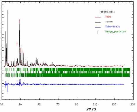
The ab initio crystal structure determination from powder XRD data was carried out with Fullprof [5] and EXPO [6]. Firstly, the structure factor amplitudes were extracted by the Le Bail method [7] from the powder pattern in Fullprof and, subsequently, the structures were solved by direct methods with Expo. Although all atoms were located at once, re-labelling of atoms was required, coupled with changes in bond distances and bond angles. This modus operandi was alternated with least-squares refinements. The coordinates of atoms obtained from direct methods were used in the Rietveld refinement of the structure by the FullProf programme. The final profile analysis refinement was carried out in the range 12.00 – 140º 2q.. The final profile fit is shown in Fig. 1. The structure of K2ZrSi3O9.2H2O are presented in Figure 2.
|
|
|
|
Fig. 1: Observed, calculated, and difference X-ray diffraction patterns of K2ZrSi3O9.2H2O |
Fig. 2: Projection of the structure of K2ZrSi3O9.2H2O |
[1] Z. Lin et al, J Phys.Chem, 1999, 103, 957-963
[2] C. Dong, J. Appl. Cryst., 1999, 32, 838.
[3] P.E. Werner, L. Eriksson, M. Westdahl, J. Appl. Cryst., 1985, 18, 367.
[4] J. Laugier, B. Bochu, Programme d'affinement des paramètres de maille à partir d'un diagramme de poudre, Laboratoire des Matériaux et du Génie Physique, Ecole Nationale Supérieure de Physique de Grenoble (INPG), Domaine Universitaire BP 46, 38402 Saint Martin d'Hères.
[5] J. Rodriguez-Carvajal, FULLPROF Program for Rietveld Refinement and Pattern Matching Analysis; Abstracts of the Satellite Meeting on Powder Diffraction of the XVth Congress of the International Union of Crystallography, Toulouse, France, 1990, p. 127.
[6] A. Altomare, M.C. Burla, M. Carmalli, B. Carrozzini, G.L. Cascarano, C. Giacovazzo, A. Guagliardi, A. Moliterni, G. Polidori, R. Rizzi, J. Appl. Cryst., 1999, 32, 339.
[7] A. Le Bail, H. Duroy, J. L. Fourquet, Math. Res. Bull., 1988, 23, 447.