g’-Fe4N formation upon annealing e-Fe3N: A Powder Diffraction study using Synchrotron Radiation
T. Liapina1,
A. Leineweber1, E. J. Mittemeijer1, M. Knapp2,
C. Baehtz2, Z.Q. Liu3, K. Mitsuishi3, and K.
Furuya3
1Max Planck Institute for Metals Research,
Heisenbergstraße 3, 70569 Stuttgart, Germany.
2Institute for Materials Science, Darmstadt
University of Technology, Petersenstr. 23, 64287 Darmstadt, Germany.
3Nanomaterials Laboratory, National Institute for Materials Science, Tsukuba 305-0003, Japan
The investigation
of iron nitrides is largely motivated due to their role in metallurgy and also
their possible potential as magnetic recording materials. The most important
iron nitride phases are e-FeNy (y = 0.22-0.49) and
g’-Fe4N. The crystal structure of e-FeNy is based on a hcp arrangement
of Fe whereas in g’-Fe4N the arrangement of
Fe is fcc. In both cases N occupies octahedral interstitial sites. Most studies
on iron nitrides were performed on compound layers generated on the surfaces of
iron or steel. The iron nitrides in such layers contain strong nitrogen
concentration gradients caused by the inward diffusion of nitrogen during
nitriding. Homogeneous iron nitrides can be prepared e.g. by nitriding of thin
iron foils or powders for moderate times. Such iron nitride powders are well
suited for structural analysis by powder diffraction methods. X-ray powder
diffraction is able to determine quantitatively very small changes in
composition [1] and the presence of compositional inhomogeneities [2].
Previous
experiments on the behaviour of e-iron nitride powders upon annealing
showed [3] that at 350°C e-FeN0.33 forms
precipitates of g’-Fe4N, which leads to an
enrichment of the remaining e-phase with nitrogen (final
composition FeN0.36), in accordance with the phase diagram (Figure
1) and as also observed for bulk specimens [4]. However, details of the
transformation mechanism are unknown at present.
|
|
|
|
Figure 1. Simplified section of the Fe-N phase diagram. y is the concentration of the initial e-phase after nitriding, and yeq is the concentration of the e-phase in equilibrium with the precipitated g’ at the applied annealing temperature. |
Figure 2. 113 reflection of e-FeNy as obtained by powder diffraction using
synchrotron radiation: original sample (after nitriding) and sample annealed at 673
K for 1 hour, 1 and 3 days, respectively. The shifts of the reflections are
mainly caused by an increase in nitrogen content. |

In this paper
high resolution powder diffraction data obtained using synchrotron radiation
(B2 at HASYLAB, Hamburg, l = 1.13985
Å) on original
and heat treated (up to 3 d at 360°C and 400°C, respectively) FeN0.33
powder (particle size 2-5 mm) are presented. The data reveal
with increasing annealing time the formation of g’-Fe4N and a gradual
increase of the N content in e-FeNy, as
evidenced by the emergence of reflections of g’-Fe4N and shifts of the
reflections of the present e-FeNy (Figure 2).
Furthermore, anisotropic and asymmetric diffraction-line broadening of the e reflections
was observed.
The narrowest e reflections
are observed for the sample annealed for 3 d at 400°C, in which apparently
equilibrium between e and g’ has been reached already.
An analysis of
the dependence on the directions of the diffraction vector of the broadening
and the asymmetry of the reflections of the different samples shows an
important contribution due to inhomogeneities (local variations in the N
content [4]). These inhomogeneities can
be reduced by further annealing leading to their virtual disappearance after 3
d at 400°C.
Transmission
electron microscopy performed on the powder particles (Figure 3) reveals that g’-Fe4N grains are formed only in
relatively few powder particles. These observations together with the finding the
narrow reflections in the powder diffraction patterns taken after long-term
annealing exclude the presence of large local composition variations (e.g. from
particle to particle), it can be concluded that the nitrogen atoms can move
from powder particle to powder particle. This can only occur via direct contact
of the mainly spherical particles, since N transport via the gas phase can be
excluded: loss of N2 to the atmosphere is well known to be fully
irreversible [5].
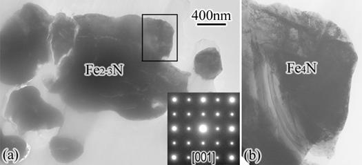
Figure 3. (a) Bright field image of some particles in an e-FeNy + g’-Fe4N sample. Inset is an g’-Fe4N diffraction pattern ([001] zone) of the top right area of the central Fe2-3N (FeNy) particle. (b) The Fe4N grain outlined in (a).
1. T. Liapina, A. Leineweber, E. J. Mittemeijer, W. Kockelmann, Acta Mater. 52 (2004) 173-180.
2. A. Leineweber, E.J. Mittemeijer, J. Appl. Crystallogr. 37 (2004) 123-135.
3. A. Leineweber, PhD thesis, University of Dortmund (1999).
4. T. Liapina, A. Leineweber, E.J.
Mittemeijer, Scr. Mater. 48 (2003) 1643-1648.
5. E. Lehrer, Z.
Elektrochem. 36 (1930) 383-392.