The Egg-Shell Microstructure Studied by Powder Diffraction
L. Dobiášová†1, R. Kužel1,
H. Šíchová1, J. Kopeček2
1Faculty of Mathematics and Physics, Charles University, Ke Karlovu 5, 121 16 Praha 2
2Institute of Physics, Academy of Sciences of the Czech Republic
† in memoriam
In last years, traditional technique of powder diffraction known mainly to materials scientists, physicists, chemists, mineralogists is also applied to biological materials. First powder diffraction studies of protein structures has appeared [1, 2]. However, powder diffraction is known also as a suitable tools for studies of the so-called real structure of materials. In present work, we have tried to perform more complete diffraction analysis of different egg-shells.
The biological function of the egg-shell is a chamber for embryonic development and from which the chick is able to emerge at the appropriate time. The requirements of the table egg industry are different. The industry sustains economic loss from cracked eggs and some of the cracking can be attributed to the deficiencies in the egg-shell structure. This is one of the reasons why the attention to egg-shell is devoted [3-5].
The egg-shell consists of several mutually through-growing layers of CaCO3. The inner most layer – mamilary layer ( ~100 µm) grows on the outer egg membrane and creates the base on which the palisade layer constitutes the thickest part (~ 200 µm) of the egg-shell. The top layer is the vertical layer (~ 5-8 µm) covered by the organic cuticle. Different kinds of hen´s and bird´s egg-shells in the powder form or as a whole from both sides of the shell were examined by powder diffractometry and film back-reflection method. The powder patterns were evaluated by the fitting of diffraction profiles with the Pearson VII function.
The lattice parameters, peak intensities and profile broadening were analysed. At the Bragg-Brentano set ting (2Q = 40°) the Cu radiation penetrates approximately into the 9 µm of the egg-shell, so the measurements from the inner and outer shell surface can give evidence of the mamilary and palisade layer, respectively. The results obtained on egg-shells of very different origins shown no significant differences in lattice parameters that correspond well to the PDF-2 values. The patterns contained only basic phase CaCO3 (space group no. 167: R-3c) with a small addition of magnesium (0.3 wt. % , determined by atomic absorption). Diffraction patterns of powders obtained from all the eggs investigated correspond very well to the pattern of standard CaCO3. The correspondence is very good including intensities. The patterns obtained from egg-shell powders are also very similar to the standard pattern, regardless larger line broadening.
However, there are differences between powders and both sides of the shells. For inner shell surfaces, the intensities are only slightly different than in powders (including standard one) but there is significant line broadening indicating fluctuations of lattice spacings (the mean local strain of about 0.2 %). On the other hand, for outer shell surfaces, there is much smaller broadening of lines, similar to powders, but significant changes of intensities indicating the (00l) textures of grains. This is also an evidence of presence of two basic layers, structurally very different – mamilary and palisade. The meaning of crystallographic texture has been emphasized [3, 4]. It was stated that the breaking strength of the egg shell is inversely related to the degree of calcite orientation and conversely, reduced strength in the egg shell from aged hens coincides with a high variability of texture [3].
As a general conclusion and amazing fact, we can say that any differences of XRD parameters (for inorganic – calcite part) between the eggs of very different origin are not significant. So that their microstructure and composition, as they can be seen by XRD, are the same. All the shells investigated exhibited strong texture from outside and no texture from inside (Fig. 1). This agrees with the SEM pictures (Fig. 2) and known fact that from smaller more or less isotropical grains larger columnar grains are developed to the outer side. This work was an attempt for non-traditional application of powder diffraction with the aim to show that the method may be helpful also for biologists. Not only because of the phase analysis but also for the study of nanostructure of inorganic crystalline phases in biological objects. This is closely related to the overall microstructure strongly influenced by proteins taking part in its creation. The egg-shell matrix proteins influences the process of crystal growth by controlling size, shape and orientation of calcite crystals. The formation of avian eggs belongs to most rapid mineralization processes known.
1. R. B. Von Dreele, "Combined Rietveld and Stereochemical Restraint Refinement of a Protein Crystal Structure," Journal of Applied Crystallography 32, 1084-1089 (1999).
2. http://lansce.lanl.gov/re search/vondreele.html.
3. Y. Nys, J. Gautron, M. D. McKee, J. M. Garcia-Ruiz, M.
T. Hincke, Biochemical and functional characterisation of egg shell
matrix proteins in hens. World's Poultry Science Journal 57 (2001) 401-413.
4. R.M.G. Hamilton, The Microstructure of the Hen's Egg Shell - A short review, Food Microstructure 5 (1986), 99-110.
5. P. Hunton, Understanding the architecture of the egg
shell, World's
Poultry Science Journal 51 (1995)
141-147. Materials Structure, vol.
10, number 1 (2003) 39.
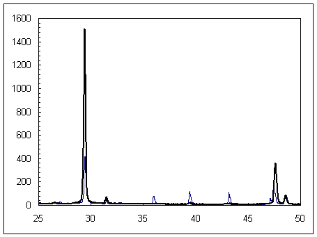
Figure 1. A
typical part of the diffraction pattern of the egg-shell (CaCO3) - from the inner (thin line) and outer side
(thick line), respectively.
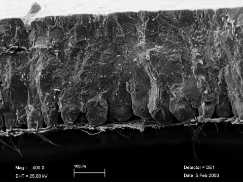
Figure 2. SEM
picture of egg-shell. Outer side is on the bottom.