Model of
disorder and structure refinement of illite
S. Ferrari1,*, A.F. Gualtieri1, M. Leoni2
1 University of Modena and Reggio
Emilia, Department of Earth Sciences, Via S.Eufemia, 19, 41100, Modena, Italy.
2 University of Trento, Department of
Materials Engineering and Industrial Technologies, Via Mesiano, 77, 38050,
Trento, Italy
* e-mail: ferrari.simone@unimore.it
The term illite, introduced by Grim in 1937 [1], refers to an aluminum-potassium mica-like, non-expanding, dioctahedral mineral, present in the clay fraction (under 4 μm). Together with kaolinite, clorite and illite-smectite mixed-layers (I-S), illite is one of the four major constituents of argillaceous sedimentary rocks. Illite generally crystallizes in the monoclinic system, like other micas. Its structure is very similar to 2:1 mica structure, with two tetrahedral sheets and one octahedral between, to build up the T-O-T sheet [2].
According to Rosenberg [3], an approximate formula for illite can be written as:
K0.88Al2(Si3.12Al0.88)O10(OH)2
deduced both by experimental studies [4, 5], and by studies on natural materials [6] With respect to muscovite, in illite there is a lower K content, due to the lower substitution of Si with Al in the tetrahedral site.
Illite presents extensive structure disorder likely due to
shifts and rotations between the layers: this contributes to the anisotropic
broadening of peaks width in the diffraction pattern. One of the major problem
concerning illite studies is finding of a pure sample. The sample utilized in
this work is pure and occurs in a mine from the Tokaji mountains region,
Hungary. The aim of this study is to investigate the structure model and
structure disorder of this pure illite sample. In order to do this, DIFFaX [7]
has been utilized for the preliminary simulation of the XRPD pattern, and
DIFFaX+ [8] for the refinement of the structure. The result obtained with
DIFFaX+ will be cross checked with WILDFIRE© [9]. Figure 1 shows the observed XRPD pattern of the pure
illite sample.
We are actually in the process to test a number of different models and to obtain the best results for our sample.
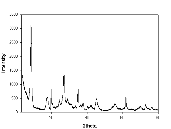
Figure 1. Observed pattern of the Hungarian illite in the range 3-80 °2θ.
1. Grim, R.E., Bray, R.H. e Bradley, W.F. (1937) American Mineralogist, 22, 813-829.
2. Brigatti, M.F. e Guggenheim, S. (2002) Mineralogical Society of America Reviews in Mineralogy, 46, 1-97.
3. Rosenberg, P.E. (2002) American Mineralogist, 87, 103-107.
4. Yates, D.M. e Rosenberg, P.E. (1996) Geochimica et Cosmochimica Acta, 60, 1873-1883.
5. Yates, D.M. e Rosenberg, P.E. (1997) Geochimica et Cosmochimica Acta, 61, 3135-3144.
6. Inoue, A., Kohyama, N, Kitagawa, R. e Watanabe, T. (1987) Clays and Clay Minerals, 35, 111-120.
7. Treacy, M.M.J., Newsam, J.M. and Deem, M.W. (1991) Proc.R.Soc.Lond.A, 433, 499-520.
8. Leoni, M., Gualtieri, A.F. and Roveri, N. (2004) Journal of Applied Crystallography, 37, 166-173.
9. Reynolds, R.C. (1993) CMS Workshop lectures Volume 5: Computer Applications to X-Ray Powder Diffraction Analysis of Clay Minerals.