X-ray powder
microdiffraction in the analysis of art works
Šímová V.1, Bezdička P1.,
Hradilová J.2, Bayerová T.3, Hradil D.1
1Institute of Inorganic Chemistry AS
CR, 250 68 Řež, Czech Republic.
2Academy of Arts, U Akademie 4, 172
22 Praha 7, Czech Republic
3University of Applied Arts,
Salzgries 14, A-1010 Vienna, Austria
e-mail:
veronika@iic.cas.cz
Analysis of samples smaller than 1 mm has always been a problem for
powder diffractometry. X-ray powder microdiffraction is a new laboratory technique,
extremely quickly developing in last three years, and becoming available mainly
due to hardware development of conventional powder diffractometers. Common
X-ray tube and monocapillary producing a quasiparallel beam combined with a
sensitive/fast solid-state detector enable direct phase analysis from 0.1 mm
spots using a traditional diffractometer in less than 1 day. It is possible to
analyse a selected place on a solid sample with irregular shape.
The
microdiffraction was applied to analyses of colour layers of art works, e.g.
canvas and wall painting and polychromy on wood. Materials research of colour
layers helps to date artworks and identify repaints, to study painting
techniques of different authors and historical periods. Powder diffraction is
extremely well suited to distinguish inorganic pigments of different natural
provenance and to reveal secondary mineralization deteriorating artworks.
The fragments
with characteristic dimensions 1x1x0.2 mm were examined by a combination of
SEM/EDX and optical microscopy (fig. 1). Microdiffraction was used to confirm
the presence of mineral pigments assumed indirectly on the base of their
elemental composition, to identify the minerals in earthy pigments and, in the
case of wall paintings, to describe the phase composition of secondary salt
efflorescences.
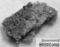
Figure 1: Optical microphotograph of fragment of Funeral
Crown of Charles IV
In the case of
polychrome funeral insignias from the Prague castle tombs, the microdiffraction
was able to directly identify the structural forms and a method of mediaeval
preparation of two different lead-tin yellows used to paint the royal crown of
Charles IV. (Fig. 2). In the first fragment, the lead-tin yellow of type I
(Pb2SnO4) was identified. In the second fragment, SEM/EDX
indicated an excess of tin and the presence of silicon, which is typical for
the lead-tin yellow of type II, i.e.
type I recrystallized in the flux with SiO2. However, type II was
not confirmed by powder X-ray microdiffraction, the excess Sn was found as
cassiterite and Si as quartz.
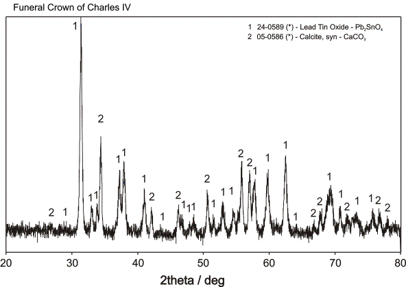
Figure 2: Diffractogram of fragment of Funeral
Crown of Charles IV
In the case of Renaissance
wall paintings from Malta, the microdiffraction was able to distinguish among
different crystalline salts built by common elements (Ca, Mg, K, S, O, N, H)
and to indicate those formed as a result of previous restoration of the
painting. (Fig. 3).
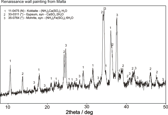
Figure 3: Diffractogram of fragment of Renaissance wall painting from Malta
Based on microdiffraction measurements, bole grounds of Baroque paintings were distinguished according to their mineralogy: kaolinite, illite, smectites, hematite (a-Fe2O3), goethite (a-FeOOH), and jarosite (basic ferric sulphate) were found as a result of different natural genesis and further treatment of earthy pigments used.
Acknowledgements:
V. Šímová
was supported by a Grant Agency of AS CR (project number B1032401), J. Hradilová,
D. Hradil and P. Bezdička acknowledge a support by Grant Agency of CR
(project number 203/04/2091). Financial support by the Ministry of Education of
CR (project number LN00A028) is also gratefully acknowledged.