Structure of the quaternary alloy Zn0.6Mn0.4In2S4 by Synchrotron Powder Diffraction and Electron Transmission Microscopy
Rosario Ávila-Godoy1, Asiloé J. Mora2, Dwight Acosta-Najarro3, Andrew N. Fitch4,
Gerzon E. Delgado2, Andrés E. Mora1,5,
John Steeds5
1Departamento de Física,
Universidad de Los Andes, Mérida-Venezuela
2Departamento de Química,
Universidad de Los Andes, Mérida-Venezuela
3Instituto de Física, Universidad
Nacional Autónoma de México, DF, México
4European Synchrotron Radiation Facility, Grenoble Cedex, France
5University of Bristol, UK
When the solid solution of
Zn0.6Mn0.4In2S4 is formed, the
material departs from the stoichiometry of the parent compound ZnIn2S4
[1], a defect-type layered semiconductor that has octahedral and
tetrahedral sites in which the cations can be accommodated. Therefore, it is
possible that some cationic positions lose their ![]() point symmetry, because it becomes necessary to
accommodate different proportions of the Zn, In and Mn cations in these sites.
Hence, a change of crystalline symmetry from R
point symmetry, because it becomes necessary to
accommodate different proportions of the Zn, In and Mn cations in these sites.
Hence, a change of crystalline symmetry from R![]() m
to R3m is possible. Also, taking into
account the ionic size, oxidation state and coordination number of Mn2+,
it is probable that the magnetic ions occupys either octahedral or tetrahedral
positions. Optical and magnetic measurements are contradictory in this matter
[2]. The objective of the present work was to determine the structure of the
quaternary alloy Zn0.6Mn0.4In2S4,
and to locate in a precise way the positions of the Mn2+ ion in the
crystalline cell. This was accomplished by means of two complementary
techniques: X-ray powder diffraction using synchrotron radiation and Electron
Transmission Microscopy techniques, such as High Resolution Microscopy (HRM)
and Convergent Beam Electron Diffraction (CBED).
m
to R3m is possible. Also, taking into
account the ionic size, oxidation state and coordination number of Mn2+,
it is probable that the magnetic ions occupys either octahedral or tetrahedral
positions. Optical and magnetic measurements are contradictory in this matter
[2]. The objective of the present work was to determine the structure of the
quaternary alloy Zn0.6Mn0.4In2S4,
and to locate in a precise way the positions of the Mn2+ ion in the
crystalline cell. This was accomplished by means of two complementary
techniques: X-ray powder diffraction using synchrotron radiation and Electron
Transmission Microscopy techniques, such as High Resolution Microscopy (HRM)
and Convergent Beam Electron Diffraction (CBED).
In spite of collecting the diffraction data in a spinning borosilicate capillary with the powder diffractometer of beamline ID31, ESRF, prefered orientation along the [001] direction due to the crystal morphology was present. However, the presence of reflection 006 at 14.3º 2q, implied that the structure could be non-centrosymmetric R3m, and Rietveld refinements using different cationic arrengements were performed. A model in which the tetrahedral sites were occupied by a random distribution of Zn, Mn and In atoms, but with local 3m symmetry, gave the best results. The Rietveld refinement of this model led to figures of merit: Rwp = 9.9%, Rp= 9.2%, χ2 = 11.21 and R(F2) = 0.1146. The final Rietveld plot showing the observed, calculated and difference patterns of the Zn0.6Mn0.4In2S4 is shown in Figure 1. Selected Area Electron Diffraction (SAED) patterns and High Resolution Micrography along [001] showed the rhombohedral configuration (Figure 2a). From CBED patterns perpendicular to [001] the 6mm symmetry breaking to the 3m symmetry associated to the R3m space group can be observed in Figure 2b.
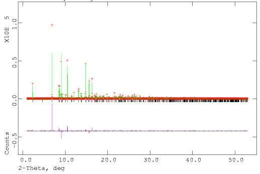
Fig. 1. Final Rietveld plot showing the observed, calculated and difference
patterns of the Zn0.6Mn0.4In2S4
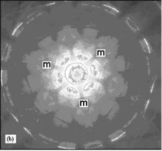
Fig. 2: (a)
High-resolution micrograph, (b) CBED pattern of Zn0.6Mn0.4In2S4 along [001] showing the 3m symmetry that validate the R3m space group.
Acknowledgments
W. Giriat and A. López-Rivera, which kindly prepared the
samples. CDCHT-ULA, FONACIT (Lab-97000821) and ESRF (France).
1. S.A. López, A.J. Mora, D. Acosta-Najarro, A.V. Rivera, and R. Ávila-Godoy (2001), Semicond. Sci. Technol., 16, 367; F. Lappe, A. Niggli, R. Nitsche, and J.G. White (1962). Z. Kristallogr., 117, 146 ; N. Berand, and K. J. Range (1994). J. Alloys Comp., 205, 295.
2. S.A. López-Rivera, L. Martínez, W. Giriat, and F. Medina (1995). Semicon. Sci, Technol., 10, 645; B. Fontal, S.A. López-Rivera, L. Martínez, and W. Giriat (1996). Semicond. Sci. Technol., 11, 1056; C. Pineda, J.M. Martín, and S.A. López-Rivera (1998). Rev. Mex. Fís., 44, 3, 224; F. Palacio, J. Campo, V. Sagredo, G. Attolini, and C. Pelosi (1995). J. Magn. Magn. Mat., 140-144, 2023.