Effect of hydrostatic pressure
on the gamma-polymorph of glycine: a phase transition
E.V. Boldyreva1,2, S.N. Ivashevskaya1,3, H. Sowa4, H. Ahsbahs4, H.-P. Weber5,6
1Novosibirsk State University, Research and
Education Center “MDEST”, Department of Solid State Chemistry, Pirogova, 2, Novosibirsk, 90, 630090 Russia
2Institute of Solid State Chemistry and
Mechanochemistry, Russian Academy of Sciences, Kutateladze, 18 Novosibirsk, 128, 630128 Russia
3Institute of Geology Karelian Scientific Center
Russian Academy of Sciences, Pushkinskaya, 11, Petrozavodsk, 185610 Russia
4Philipps-Universitat Marburg/Lahn, Institute of Mineralogie, Hans-Meerwein Strasse, D-35032, Marburg/Lahn, Germany
5European Synchrotron Radiation Facility, Swiss-Norwegian Beamlines, PO Box 220, F-38043, Grenoble CEDEX, France
6Institut de Cristallographie, Universite de Lausanne, CH-1015 Lausanne , Switzerland
Introduction
While the majority of crystal structures of organic molecules have been determined at normal pressure conditions, there is a great demand for the observations of structural changes that occur in organic solids in response to high pressure.
Among molecular organic crystals, those of amino acids attract special attention – as biomimetics, as solid drugs, as materials for molecular electronics, as systems important for geo- and cosmochemistry.
The hot topics of the research are:
- the search of high-pressure polymorphs of amino acids,
- the studies of the anisotropy of pressure-induced structural distortion not accompanied by a phase transition.
Experimental
X-ray
powder diffraction patterns were measured in transmission mode. A
monochromatized synchrotron radiation source of the Swiss-Norwegian Beam Line
at ESRF was used (l
= 0.71950 A) for detailed studies, since glycine is a poor diffractor (having
only light N, O, C and H atoms). Diffraction patterns were registered with a
MAR345 image plate detector (pixel size 0.15 mm, 2300 x 2300 pixels in image,
maximum resolution 1.105 A, maximum 2q = 36.942 deg.). The frames were measured with exposing
time equal to 900 - 3600 seconds, with Df = 0.03 degrees. The distance
crystal-detector, the beam center position, the tilt angle and the tilt plane
rotation angle were refined using a Si standard put at a diamond anvil (DAC). A
methanol-ethanol mixture was used as a pressure-transmitting medium [1]. It was
specially dried to have no traces of water, because even traces of water are
known to influence on the polymorphic transformations in glycine. The sample in
the DAC was centered with respect to the beam very carefully, so that no
reflections from steel gasket could be observed in the measured diffraction
pattern.
Fit2D
program [2] was used for processing diffraction data measured with the
synchrotron source (calibration, masking, integration).
The
unit cell dimensions were determined with the indexing program TREOR [3] with M19=
22, F19 = 48 (0.009163, 44) using the first 19 peak positions. The
structure was solved by the grid search procedure [4] and refined with the use
of bond restraints by the MRIA program [5]. The strength of restraints was a
function of interatomic separation and for intramolecular bond lengths
corresponds to an r.m.s. deviation of 0.03 A. H atoms were placed in
geometrically calculated positions and allowed to refine using bond restraints
with a common isotropic displacement parameter Uiso fixed to 0.05 A2.
PowderCell
[6] was used for structure analysis and graphic representation.
Results and discussion
At 2.74 GPa the reflections of a new phase could be observed, although g-polymorph was still the major component at this pressure. The new high-pressure phase was present as the main component in the pressure range 4.17 – 7.85 GPa, but even at 7.85 GPa the peaks of the low-pressure phase (g-glycine) were still present in the diffraction patterns.
The structure of a new high-pressure polymorph was solved in the Pn (No 7) space group (a = 5.379(1)A, b = 5.557(1)A, c=4.780(1)A, b = 118.25(1)o, V = 125.86(4)A3, Z = 4). The packing of zwitter-ions in the high-pressure polymorph turned out to be essentially different from that in the original g-polymorph (P31), but similar in many respects to packing of zwitters-ions in the other two previously known polymorphs of glycine – a- (P21/n) and b- (P21) forms. [7]. In the g-polymorph zwitter-ions are linked via hydrogen bonds in a three-dimensional network based on helical chains. In the new high-pressure polymorph the zwitter-ions are rearranged to give specific layers.
The structure of an individual layer in the high-pressure polymorph is similar in many respects to the structures of individual layers in the a- and b- forms, but the way how the layers are stacked is essentially different: the layers in the high-pressure polymorph are double, as in the a-form, but the individual layers in the double layer are related not by inversion, as in the b-form, but by a glide plane. The pressure-induced polymorphic transformation in the g-polymorph can be compared to a change in the secondary structure of a polypeptide chain from a helix into a layer.
A high resolution, synchrotron X-ray pattern of the glycine high-pressure modification and a difference between the measured and calculated profiles are shown on Figure 1. Atomic coordinates and isotropic displacement parameters are in the Table 1.
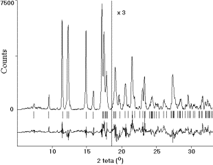
Figure 1
Table 1
|
atom |
x |
y |
z |
Uiso (A2) |
|
O1 |
0.9533 |
0.806(2) |
0.2824 |
0.018(1) |
|
O2 |
0.308(2) |
0.6525(14) |
0.767(2) |
0.018(1) |
|
N1 |
0.780(3) |
0.724(2) |
0.734(2) |
0.018(1) |
|
C1 |
0.224(3) |
0.763(3) |
0.519(2) |
0.018(1) |
|
C2 |
0.484(3) |
0.853(2) |
0.496(3) |
0.018(1) |
|
H1 |
0.516 |
0.022 |
0.549 |
0.051 |
|
H2 |
0.433 |
0.825 |
0.294 |
0.051 |
|
H3 |
0.753 |
0.560 |
0.690 |
0.051 |
|
H4 |
0.945 |
0.782 |
0.708 |
0.051 |
|
H5 |
0.850 |
0.756 |
0.943 |
0.051 |
On decompression, the high-pressure phase did not disappear completely even at ambient pressure. At 3.27 GPa the amount of the initial g-polymorph increased considerably. Besides, some additional lines appeared that could not be assigned either to the new high-pressure polymorph, or to the original g-form.
Acknowledgement
The study was supported by RFBR (grant 02-03-33358),
the BRHE-Program (grant NO-008-XI), Russian Ministry of Education (grants
Ч0069 of the “Integration”-Program; ЗН-67-01; and
ур.05.01.021 of the Program “Universities of Russia”), and the
National Science Support Foundation for EB (Program “Young Professors”). The
diffraction experiment using synchrotron radiation was carried out at the
Swiss-Norwegian Beamline at ESRF (Grenoble), experiments 01-02-656 и
01-02-671). The authors are grateful to V.V. Chernyshev and V.B. Zlokazov for
their assistance in the use of MRIA program and valuable advice.
[1] G. J. Piermarini, S. Block, J. D. Barnett, J. Appl. Phys. 44 (1973) 5377-5382.
[2] A. Hammersley,
Version V11.012, hammersley@esrf.fr.
[3]
P.-E. Werner, L. Eriksson, M. Westdahl, J. Appl. Cryst. 18 (1985)
367-370.
[4]
V.V. Chernyshev, H. Schenk, Z. Kristallogr. 213 (1998) 1-3.
[5]
V.B. Zlokazov, V.V. Chernyshev, J. Appl. Cryst. 25 (1992) 447-451.
[6]
W. Kraus, G. Nolze, PowderCell for Windows, Vers. 2.3, http://www.bam.de/a_v/v_1/powder/e_cell.html.
[7] E. Boldyreva, H. Ahsbahs, H.-P. Weber, Z. Kristallogr. 218 (2003) 231-236.