(b / a)8 BARREL ENZYMES OF THE HISTIDINE BIOSYNTHESIS PATHWAY
Dietmar A. Lang1,
Galya Teplyakova1,
Ralf Thoma2,
Kasper Kirschner2,
Reinhard Sterner2,3
and Matthias Wilmanns1
1EMBL Hamburg Outstation c/o DESY,
Notkestrasse 85, D-22603 Hamburg, Germany
2Biozentrum Basel,
Klingelbergstrasse 70, CH-4056 Basel,Switzerland;
3Institute for Microbiology and
Genetics, University of Göttingen, Grissbachstrasse 8, D-37077
Göttingen, Germany.
Histidine biosynthesis was presumably assembled prior to the diversification of bacteria, archaea and eucaryotes [1]. In Escherichia coli the unbranched pathway that requires ten enzymatic reactions is coded by eight genes [2]. In shaping the pathway, evolutionary gene duplication might have played a major role and it was postulated that two genes, hisA and hisF ,have evolved by internal gene duplication [3]. The gene products of hisA and hisF, phosphoribosyl-formino-5-amino-1-phosphoribosyl-4 imidazole carboxamide isomerase and the cyclase subunit of the imidazoleglycerol phosphate synthase, were predicted to be folded as (b / a)8 barrel proteins using threading methods [5] and sequence comparison techniques [6].
To shed light into the evolutionary enzyme development of this pathway we have started 3D structure analysis of the hisA (241 residues) and hisF (253 residues) gene products from the thermophilic strain Thermotoga maritima .
We have obtained a monoclinic crystal form of HisF that diffracts to better than 1.0 Å resolution. Its 3D structure has been determined from a MAD data set using selenomethionine incorporated crystals. The data were collected on the tuneable wiggler beam line BW7A at the EMBL Hamburg Outstation. The positions of the five selenium sites were determined from Difference Patterson maps. The resulting phases were refined and extended using the programs SHARP and ARP. The refinement of this structure is almost completed using a native date set to 1.4 Å resolution (current R-factor, 20 %; current R-free, 22 %).
Four different crystal forms of the hisA gene product have been obtained. A native data set to 2.4 Å of a monoclinic crystal form has been collected on the high intensity BW7B wiggler beam line of the EMBL Hamburg Outstation so far. We are planning to solve this structure using a MAD data set of the same crystal form with selenomethionine incorporated HisA.
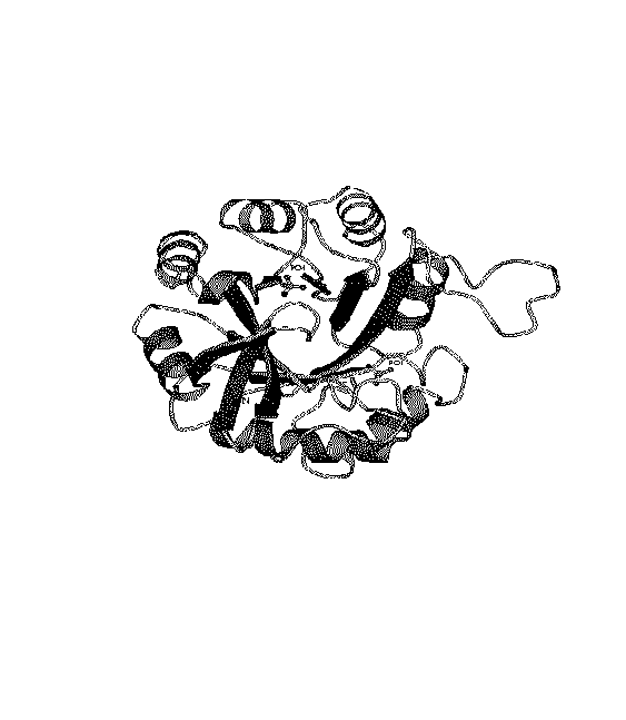
The structure of the cyclase subunit of the imidazoleglycerol phosphate synthase (HisF; figure 1) reveals some features which were already predicted in the past:
Figure 1: Ribbon of the HisF structure.
The view is along the barrel axis from the C-terminal side of the
central b-barrel. The two phosphate sites are included in
ball-and-stick representation.