STRUCTURE OF CYANAMIDONITRATE-COPPER(II) COMPLEXES OF IMIDAZOLE LIGANDS; EQUATORIAL-AXIAL CORRELATION
Jozef Kozisek, Jess Garca Daz, Maria Hvastijova, Martin Breza and Ingrid Svoboda1
Faculty of Chemical Technology, Slovak
University of Technology, 812 37 Bratislava, Slovakia
e-mail : kozisek@cvt.stuba.sk
1Fachbereich
Materialwissenschaft, Technische Universitt Darmstadt, D-642 87
Darmstadt, Germany
A well-known equatorial-axial correlation [1] in hexacoordinate complex compounds is obeyed by small, but significant changes in equatorial and axial bond lengths. This phenomenon has quite comprehensible reason: the limited bonding ability of the central atom. Bonds-strengthening in equatorial planes is compensated by bonds-weakening in axial direction and vice versa. Copper(II) complexes with CuO6, CuN4O2 and CuN6 chromophores [2] represent the most common and the most numerous examples of this phenomenon. This correlation may be easily confirmed using the Cambridge Crystallographic Database. However, the question on the reason for stronger or weaker bonding in the selected direction is not completely answered yet. There are more factors which could influence this phenomenon:
a) the s- donor bonding properties of the ligand
b) the p-donor or p-acceptor bonding properties of the ligand
c) the basicity of the ligand (especially the donor atom)
d) the hydrogen bonds and other molecular interactions
e) another chemical properties of the ligand's donor atom
Common treatments to this problem suppose the dominant role of the chromophore, molecular interactions seem to be too weak to overcome it.
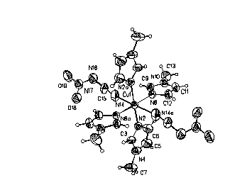
Within the systematic research of the bonding properties of non-linear pseudohalide ligand (NO2N(CN) - cyanamidonitrate) in the complexes of 3d-transition metals, the crystal structures of [Cu(1-meiz)4{NO2N(CN)}2] (1) and [Cu(5-meiz)4{NO2N(CN)}2] (2), (meiz = methylimidazole) were solved. The Cu-N distances in equatorial plane of 2.007(2) and 2.024(2), and Cu-O of 2.600(3) A in axial position for (1) agree with the values for (2) of 2.000(2), 2.014(2), and 2.683(3) A, respectively. Despite very similar molecular structure of both the compounds, there are significant differences in the molecular packing pattern of the molecules in the crystal. Distinct orientations of imidazole rings as well as of cyanamidonitrate ligands to the equatorial plane should be connected with different hydrogen bonds and molecular interactions. The angles of imidazole ring planes to the equatorial plane for (1) are 37.0(1) and 102.7(1)o, and for (2) 173.9(1) and 88.0(1)o , respectively. The angle of cyanamidonitrate plane to the equatorial plane gives 145.6(1) for (1) and differ from the value of 56.0(1)o for (2) as well.
Here the question arises what is the origin of the differences
observed: either intra-chromophore reasons based on the changes
in the electronic structure of equatorial ligands (1-meiz vs.
5-meiz) or different inter-chromophore molecular interactions
(especially hydro-gen bonds). Our attempt to explain this problem
is based on theoretical quantum-chemistry studies of electronic
structure of the chromophores using semiempirical INDO/2
(Intermediate Neglect of Differential Overlap) method for
transition metals complexes [3]. Preliminary results do not
confirm the leading role of the chromophores. Their geometry
differences should be explained by different molecular
interactions due to sterical factors based on different methyl
positions on the imidazole ring.
ORTEP drawing of the molecular structure of
[Cu(1-meiz)4{NO2N(CN)}2] with
50% thermal ellipsoids.
Crystal data [4] :
(1) C18H24N14O4Cu, Mr = 564.10, monoclinic, P21/n, lMoK = 0.71073 A, Enraf-Nonius CAD4 diffractometer; a = 7.285(1)A, b = 11.936(2)A, c = 14.752(3)A , b = 97.26(1), V = 1272.5(4)A 3, Z = 2, Dcalc = 1.472 gcm-3; blue-violet crystals, current R1 = 0.0357 for 1825 reflections with I > 4(I), wR2 = 0.1083 for all 2243 data, refinement on F2, 170 parameters (SHELX97).
(2) C18H24N14O4Cu,
Mr = 564.10, monoclinic, P -1, lMoKa = 0.71073A , Enraf-Nonius CAD4
diffractometer; a = 7.403(1)A, b = 9.292(3)A, c
= 10.556(2) A,a = 94.27(2), b = 106.89(1), g
= 112.45(2), V = 627.9(3) 3, Z = 1, Dcalc =
1.492 gcm-3; violet crystals, current R1 =
0.0401 for 1971 reflections with I > 4(I), wR2 =
0.1180 for all 2194 data, refinement on F2, 169
parameters (SHELX97).