X-RAY ANALYSIS ON A NEW SILVER COMPLEX WITH PHTALAZINE: COMPARISON OF SYNCHROTRON AND CONVENTIONAL RADIATION SOURCE
Miroslav Slouf 1,3,
Ivana Srnova 2,4,
Luca Olivi 1
e-mail: mirek@prfdec.natur.cuni.cz
1 Sincrotrone Trieste SpA, Padricano,
Trieste, Italy
2 Dipartimento di Scienze Chimiche,
universita di Trieste, Italy
3 Departement of Inorganic Chemistry,
Charles University, Prague, Czech Republic
4 Departement of Physical and
Macromolecular Chemistry, Charles University, Prague, Czech
Republic
Keywords: single crystal, synchrotron radiation
Diffraction data for a new Ag(I) complex with phtalazine (pht) were collected using both diffractometer with conventional radiation (CR) source at the University of Trieste [b] and synchrotron radiation (SR) source at HRXD beamline in Elettra [a]. Crystals used for both measurements came from the same crystallization and were the same size approximatelly. Results obtained were compared from the point of view of time and accuracy.
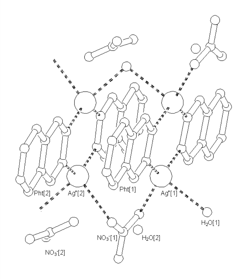
Crystal data for AgPHT = ([Ag2(H2O)(NO3)(pth)2]+ . NO3- . H2O)n (Fig.1): Mw = 635.8, triclinic, space group P-1(no.2), a = 7.473, b = 9.455, c = 14.629 A, alpha = 78,984, beta = 85.631, gamma = 79.294 degrees, V = 996A3. Dx = 2.12 g/cm3, final refinemets gave R1, wR2, GooR = 9.21%, 25.40%, 1.554 for CR and 3.81%, 10.61%, 0.939 for SR, respectively. 4330 independent reflections were collected using CR and 3322 using CR, which is 15.2(CR) and 10.8(SR) data/parameter.
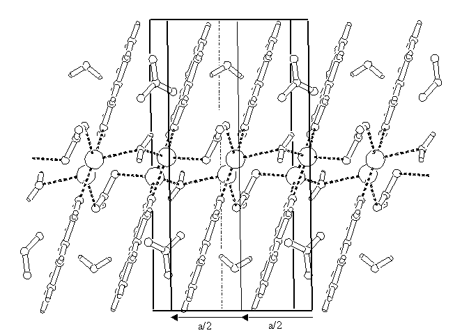
Data reduction for SR data was made using DENZO and SCALEPACK programs[1]. Structure was determined and refined by SHELXL97[2] for both CR and SR data. AgPHT is a polynuclear complex with a polymeric structure. Silver atoms are connected by phtalazine rings into infinite double chains. (Fig.2). Silver co-ordination sphere is distorted tetrahedric. There are three types of weaker intermolecular interactions in the crystal: aromatic pht-pht interactions, hydrogen bonds among H2O and NO3- molecules and weak Ag-Ag bondings. Almost all reflections with Miller index h are much weaker that those with h even. If the weaker reflections are neglected, reciprocal lattice vector a* is doubled and, as a result, real lattice vector a is half; structure is described by a smaller unit cell (Fig.2, thinner lines) and a disorder of H2O and NO3- groups. If all the reflections are included into calculation, cell is doubled (Fig.2, thicker lines) and the disorder vanishes. From that point of view AgPHT structure is a superstructure.
Standard data collection on the four cycle diffractometer with zero-dimensional detector and Mo-target X-ray tube took two days. There were some problems with catching and indexing the weaker reflections with h odd. Rotating crystal method data collection with SR source and 2D detector, on the other hand, was a routine measurement that gave stronger reflections and, subsequently, lower R-factor. Complete data set for this triclinic crystal was collected in less than two hours during the remaining time after a protein data collection. Maximum theta for CR data was 25 degrees (wavelength 0.071mn) and could have been increased easily, SR data maximum theta was 29 degrees (wavelength 0.080nm, minimum possible value for the beamline), but this was the absolute maximum for a given experiment geometry. CR data reduction required about two minutes, home-written program and a personal computer. SR data reduction took two hours, required powerful Silicon Graphics workstation and the special software.[1] So we can conclude, that standard SR data collection for "small molecule" takes just a few hours or even minutes and gives comparable or even better results than CR data collection. However, there are problems with higher theta values and data reduction is more exacting.
[1] Z. Otwinowski, W. Minor, Methods in Enzymology 1996,
276, 307
[2] G. M. Sheldrick, SHELXL-97, Universitat Gotingen, 1997