QUARTZ CRYSTAL STRUCTURE MODIFICATIONS DUE TO A SHOCK COMPRESSION AND RELEASE - DEFORMATION MECHANISM REVEALED BY RIETVELD REFINEMENT
Roman Skála
Czech Geological Survey, Geologická 6, CZ-15200 Praha 5, Czech Republic, author's e-mail address: skala@cgu.cz
Keywords: quartz, Rietveld refinement, crystal structure, deformation, shock compression
Introduction, samples, and experimental. Specific deformation features in quartz are now widely used and generally accepted as indicators of high strain due to hypervelocity impacts of extraterrestrial bodies in quartz-rich terrestrial rocks [e.g. 1, 2, 3, 4, 5, 6 and references therein]. The aim of the present paper is to demonstrate that the behavior of quartz on an atomic scale during dynamic load with extremely high strain rates (such as experienced during a meteorite impact) and shock wave release is very complex and includes significant changes of basic structure motifs.
The three experimentally shocked quartz samples, kindly provided by Bernard Champagnon (being previously investigated by the Raman spectroscopy), had been shocked at NASA Johnson Space Center, Houston, Texas, to 21.7 GPa (sample #941, N1 in [7]), 26.2 GPa (#319, N3), and 29.8 GPa (#356, N5) at 25 °C, respectively. The standard is clear quartz from Švedlár (Slovak Republic) where quartz veins are being exploited for making optical quartz glass.
Step-scanned powder data were collected in the range of 18 - 150 °2q CuKa with 0.015 °2q CuKa steps and 15 to 18 second count-time per step using a Philips X'Pert PW3710-MPD System.
Rietveld refinement of the powder data was carried out using the program FullProf [8]. Starting parameters of the crystal structure variables including space group and unit-cell choice were adopted from [9] for quartz at ambient pressure and temperature. The turning-on sequence during the Rietveld refinement followed the recommendations of [10]. Bond lengths and angles were calculated from unit-cell dimensions and fractional coordinates by the program BONDSTR [11]. To calculate polyhedral volumes, quadratic elongations, and angle variances, the program VOLCAL [12] was employed.
Distortion indices as quadratic elongation (Q.E.), bond angle variance (s 2), bond length variance (D) and distortion indices (DI) according to [13] were calculated to characterize the departure of the shape of [SiO4] tetrahedra from ideal regular geometry. Also, so-called tilt angle, f, introduced by [14], quantifying the amount of simultaneous tilting of the tetrahedra around the twofold axes perpendicular to c was calculated.
Results. The crystal structure parameters of the four quartz samples studied and corresponding distortion indices are listed in Table 1.
The unit-cell parameters and the unit-cell volume increase with the increasing peak pressure. This observation is fully compatible with data compiled in [6] and with the data published in [3]. However, the magnitude of the unit-cell volume change is considerably lower than that given in [3] though the material studied was shocked to almost identical peak pressures at the same pre-shock temperature. The observed maximum difference in cell volume is 1.6% for the sample shocked to 29.8 GPa whereas [3] gave 2.9% for a sample that experienced a shock pressure of 30 GPa.
The ratio of the unit-cell parameters c/a derived from data in [3] decreases with increasing peak pressure and the magnitude of the change of the respective parameter strongly depends on the crystallographic direction along which the pressure is applied. However, present study reveals that the c/a ratio decreases slightly only with the peak pressure applied, the difference being within a 1s range for almost all samples. This inconsistency is probably due to different geometry of the shock-recovery experiment. Langenhorst [3] used single-crystal material, which was shocked in defined crystallographic directions whereas the experimentally shocked materials used in the present study were polycrystalline.
All observed changes were very subtle for the sample #941 shocked to 21.7 GPa. More pronounced changes were found for the sample #319 that was subjected to a pressure load of 26.2 GPa. Sample #356, which was shocked to 29.8 GPa, displays the strongest changes compared to the standard, unshocked sample. The unit-cell dimensions and the derived parameters for the sample from the Meteor Crater lies between those for samples #941 and #319 but are closer to the sample that experienced shock pressure of 21.7 GPa.
In the [SiO4] tetrahedron, there are two pairs of non-equivalent Si-O bond lengths. Those are designated as d1 and d2 in this abstract, d1 being longer than d2 at ambient pressure. While the d2 distances remain almost unchanged within 1s ranges, the distance d1 decreases with increasing pressure until it becomes shorter than d2 for sample #356 at 29.8 GPa. The trend of parameter value decrease with increasing amplitude of shock load was observed also for the tetrahedral edges (O…O)T A, B, and, C, and the tetrahedral angles (O-Si-O) a and g whereas the distance (O…O)T D and angle (O-Si-O) d follow an opposite trend. The angle (O-Si-O) b remained constant. Collectively, the changes of all intra-tetrahedral parameters described above result in a pronounced decrease of the tetrahedral volume VT with increasing pressure - thus the tetrahedral volume decreases from 2.25 A3 for the unshocked quartz to below 1.95 A3 for the sample shocked to 29.8 GPa.
All inter-tetrahedral parameters increase with increasing shock pressure and thus compensate for decreased intra-tetrahedral distances and tetrahedral volumes. This agrees well with observations by Jorgensen [15] who suggested that a cooperative rigid rotation of linked tetrahedra allows compensating for the largest volume change. The increase in the angle-at-bridging-oxygen (Si-O-Si) with increasing shock comminution was confirmed earlier by e.g. Raman spectroscopy [7].
No significant correlation with peak shock pressure was observed for the distortion index DI(TO), which agrees surprisingly well with observations on statically compressed quartz samples [9, 15,16, 17]. The other distortion parameters clearly depend on the pressure load. However, the angle variance and quadratic elongation exhibit a significant deviation from the other values for sample shocked at 29.8 GPa.
Discussion. Powder diffraction has appeared to be the only method, to recover structural information from strongly shocked quartz grains because single-crystal methods has failed due to inherent mosaicism and asterism present in such materials. Using a four-circle single-crystal goniometer to study shocked material broad, diffuse reflections were obtained that did not allow meaningful data interpretation. The Rietveld method, on the contrary, provided results that allow interpretation of the mechanism(s) involved in crystal structure modification due to shock compression and post-shock pressure release.
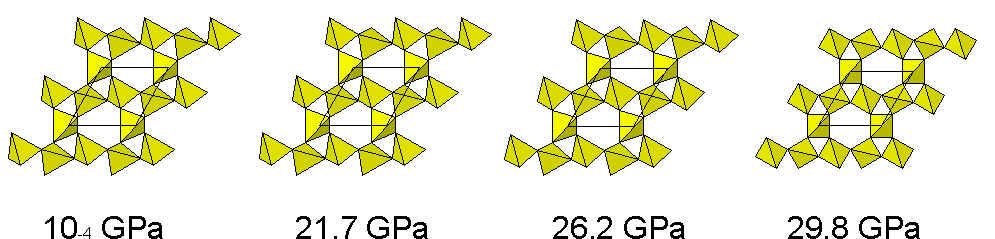
The mechanism observed involves several gradual changes. Within the quartz tetrahedron, the shortening of intra-tetrahedral distance d1 from ca 1.69 A at ambient pressure to below 1.57 A at 29.8 GPa with d2 being constant at about 1.60 A is one of the observed mechanisms. Another intra-tetrahedral change is the shortening of five of six tetrahedral edges whereas the remaining one, the (O…O) distance, increases in length with increasing pressure. When both Si-O bond lengths and (O…O) distances along tetrahedron edges change, the angles (O-Si-O) have to change as well. Three of the six angles become more acute, two remain almost unchanged, and one is more obtuse as pressure increases. Significant lengthening of inter-tetrahedral oxygen-oxygen distances, distances of tetrahedral centers (Si…Si), and opening of the angle-at-bridging-oxygen (Si-O-Si) between the two linked tetrahedra with increasing shock stress fully compensate for shortening of intra-tetrahedral parameters and obviously are responsible for the increase of unit-cell dimensions. All changes mentioned above, when applied cooperatively, lead eventually more or less to regularization of the quartz crystal structure, as it is apparent from the structure projection onto the ab plane in Fig. 1. The ditrigonal structure of a-quartz gradually changes to hexagonal-like arrangement in the sample shocked at 29.8 GPa. It is possible that extreme strain rates caused by shock stress may finally attribute to the formation of a phase with symmetry very close to the hexagonal arrangement typical of b-quartz. Another most prominent is the decrease in tilt angle for samples shocked at 26.2 and at 29.8 GPa eventually resulting in slight rotation of the tetrahedra along c. The rotation of tetrahedra is also apparent from a vertical view presented in Fig. 1.
In certain aspects, the observed modifications to quartz crystal structure are consistent with those found during single-crystal structure refinements under elevated static pressure [9, 15,16, 17]. Both the present study and the static pressure study revealed a decrease of the tetrahedral volume and a shortening of Si-O bond lengths and most tetrahedral edges with increasing pressure. However, measurements under static pressure also indicated a shortening of inter-tetrahedral distances and a decrease in the angle-at-bridging oxygen, which is not the case for the shocked samples. This fact unambiguously reveals that tetrahedra once compressed by a shock wave retain the compressed shape (maybe only slightly modified by further shock release) but the structure relaxes after shock pressure release through lengthening of inter-tetrahedral distances and opening of angle-at-bridging-oxygens. This is not the case in static compression experiments, where data are collected while the sample is subjected to pressure during measurement. Only abrupt release of shock stress is obviously capable to 'freeze' the state of shock compression within the tetrahedra while angles and distances among individual tetrahedra accommodate the structure relaxation due to pressure release, which is then expressed by the increase of the unit-cell volume. Apparently, the spirals of [SiO4] tetrahedra behave like coiled springs, allowing relatively easy contraction and expansion.
The present study constitutes a new approach using powder data collected on shocked quartz-bearing materials and provides new data, which could help us to come to a better understanding of atomic-scale mechanisms involved during shock compression and pressure release of quartz.
Acknowledgments. The author would like to express his thanks to the Grant Agency of the Czech Republic for support of the research project No. 205/95/0980. This research was undertaken in conjunction with a study dealing with shock-induced phenomena in carbonate-rich targets. Professor Bernard Champagnon kindly provided experimentally shocked quartz.
[1] R.A.F. Grieve, F. Langenhorst, and D. Stöffler, Meteoritics and Planetary Science, 31 (1996), p. 6-35.
[2] R.J. Hemley, C.T. Prewitt, and K.J. Kingma, High pressure behavior of silica. In Heaney P.J., Prewitt C.T., and Gibbs G.V., eds., Silica. Reviews in Mineralogy, 29 (1994), 41-81.
[3] F. Langenhorst, Earth and Planetary Science Letters, 128 (1994), 683-698.
[4] D. Stöffler, Fortschritte der Mineralogie, 49 (1972), 50-113.
[5] D. Stöffler, Fortschritte der Mineralogie, 51 (1974), 256-289.
[6] D. Stöffler and F. Langenhorst, Meteoritics, 29 (1994), 151-181.
[7] B. Champagnon, G. Panczer, C. Chemarin, and B. Humbert-Labeaumaz, Journal of Non-Crystalline Solids, 196 (1996), 221-226.
[8] J. Rodriguez-Carvajal, FullProf version 3.2. Rietveld, profile matching and integrated intensity refinement of X-ray and/or neutron data. A computer program. (1997)
[9] J. Glinnemann, H.E. King Jr., H. Schulz, Th. Hahn, S. J. La Placa, and F. Dacol, Zeitschift für Kristallographie, 198 (1992), 177-212.
[10] R.A. Young, Introduction to the Rietveld method. In Young, R.A. ed., The Rietveld Method, Oxford University Press, Oxford 1993, p. 1-38.
[11] J. Rodriguez-Carvajal, BONDSTR - distance, angle and bond-strength calculations. A computer program. (1995).
[12] L.W. Finger, VOLCAL - Program to calculate polyhedral volumes and distortion parameters. A computer program. (1996).
[13] W.H. Baur, Acta Crystallographica, B30 (1974), 1195-1215.
[14] H. Grimm and B. Dorner, Journal of Physics and Chemistry of Solids, 36 (1975), 407-413
[15] J.D. Jorgensen, Journal of Applied Physics, v. 49 (1978), 5473-5478.
[16] R.M. Hazen, L.W. Finger, R.J. Hemley, and H.K. Mao, Solid State Communications, 72 (1989), 507-511.
[17] L. Levien, C.T. Prewitt, and D.J. Weidner, American Mineralogist, 65 (1980), 920-930.
[18] R.M. Hazen and L.W. Finger, Comparative Crystal Chemistry - Temperature, Pressure, Composition and the Variation of Crystal Structure. John Wiley and Sons, Chichester, 1982, 231 + xv p.
Fig. 1. Projections of quartz crystal structure onto ab plane. Note gradual regularization of the crystal structure from ditrigonal symmetry up to almost hexagonal arrangement for the sample shocked to 29.8 GPa. The rotation of tetrahedra along vertical is also apparent from this view. Rhomb indicates the unit-cell.
| Table 1. crystal structures Parameters of experimentally shocked quartz materials compared to a standard | ||||
| Sample | #STD | #941 | #319 | #356 |
| Pressure [GPa] | 10-4 | 21.7 | 26.2 | 29.8 |
| Unit-cell parameters | ||||
| a [A] | 4.9126(1) | 4.9124(2) | 4.936(2) | 4.940(8) |
| c [A] | 5.4048(1) | 5.4046(2) | 5.419(1) | 5.432(6) |
| Vcell [A3] | 112.96(3) | 112.95(5) | 114.3(4) | 115(2) |
| V/V0 | 1.0000 | 0.9999 | 1.0119 | 1.0160 |
| c/a | 1.1002(1) | 1.1002(2) | 1.098(2) | 1.100(9) |
| Fractional coordinates of atoms | ||||
| Si, x | 0.4737(5) | 0.4730(6) | 0.479(1) | 0.537(3) |
| O, x | 0.4247(8) | 0.4236(8) | 0.4282(8) | 0.435(2) |
| y | 0.2990(8) | 0.2972(9) | 0.277(1) | 0.217(4) |
| z | 0.2343(8) | 0.2336(9) | 0.221(1) | 0.204(4) |
| Tetrahedral distances, d(Si-O) [A] | ||||
| d1 (2) | 1.690(4) | 1.684(5) | 1.625(5) | 1.56(2) |
| d2 (2) | 1.594(5) | 1.598(5) | 1.596(6) | 1.60(2) |
| d(Si-O) | 1.642(4) | 1.641(5) | 1.610(6) | 1.58(2) |
| Tetrahedral edges, d(O...O)T [A] | ||||
| A (2) | 2.640(6) | 2.642(7) | 2.582(7) | 2.49(3) |
| B (2) | 2.642(5) | 2.642(6) | 2.610(7) | 2.56(2) |
| C | 2.760(5) | 2.749(6) | 2.661(7) | 2.33(3) |
| D | 2.749(6) | 2.745(7) | 2.722(8) | 2.90(3) |
| d(O…O)T | 2.679(6) | 2.677(6) | 2.628(7) | 2.56(3) |
| Tetrahedral angles, (O-Si-O) [°] | ||||
| a (2) | 106.9(4) | 107.2(5) | 106.6(5) | 104(2) |
| b (2) | 107.1(4) | 107.2(4) | 108.3(5) | 108(2) |
| g | 109.5(4) | 109.4(4) | 109.9(5) | 96.3(6) |
| d | 119.1(4) | 118.4(5) | 117.1(6) | 130(2) |
| (O-Si-O) | 109.4(4) | 109.4(4) | 109.5(5) | 109(2) |
| Distance of tetrahedral centers, d(Si…Si) [A] | ||||
| d(Si…Si) (4) | 3.054(1) | 3.055(2) | 3.064(3) | 3.079(9) |
| Oxygen-oxygen distances between tetrahedra, d(O…O)IT [A] | ||||
| D1 (2) | 3.179(6) | 3.189(6) | 3.348(7) | 3.7(2) |
| D2 (1) | 3.065(6) | 3.074(7) | 3.289(8) | 3.7(3) |
| d(O...O)IT | 3.141(6) | 3.151(7) | 3.329(7) | 3.7(3) |
| Angles at bridging oxygen, (Si-O-Si) [°] | ||||
| (Si-O-Si) (4) | 136.9(2) | 137.1(2) | 144.0(3) | 154(1) |
| Distortion parameters | ||||
| tilt angle, f [°] | 22.02 | 21.87 | 17.84 | 12.34 |
| quadratic elongation, Q.E. | 1.0060 | 1.0051 | 1.0038 | 1.0334 |
| angle variance, s 2 | 23.43 | 19.95 | 15.56 | 129.86 |
| bond length variance, D(TO) | 8.42 | 6.87 | 0.84 | 1.16 |
| tetrahedral volume, VT [A3] | 2.25 | 2.25 | 2.13 | 1.93 |
| distortion index, DI(TO) | 0.029 | 0.026 | 0.009 | 0.011 |
| distortion index, DI(OO) | 0.019 | 0.017 | 0.016 | 0.046 |
| distortion index, DI(OTO) | 0.030 | 0.027 | 0.025 | 0.066 |
| Note: Estimated standard deviations of mean values (data in brackets) were calculated as s = 1/n Ssi showing an upper limit of correct standard deviations [18]. | ||||