K.-D. Liß (1), B. Waibel (2,5), R. Hock (3), A. Magerl (4), S. Köhler (1,3), A. Remhof (5)
(1) European
Synchrotron Radiation Facility, B.P. 220 F-38043
Grenoble Cédex
(2) Institut Max von Laue
- Paul Langevin, B.P. 156, F-38042 Grenoble
Cédex
(3) Mineralogisches
Institut der Universität Würzburg, Am Hubland,
D-97074 Würzburg
(4) Lehrstuhl
für Kristallographie, Universität Erlangen
Nürnberg, D-91054 Erlangen
(5) Ruhr-Universität
Bochum, D-44780 Bochum
e-mail: liss@esrf.fr
High resolution X-ray diffraction at the high energy beamline ID15A
of the European Synchrotron
Radiation Facility has been applied for the investigation of
perfect silicon crystals subjected to compressional ultrasound in
the 10 MHz range. Photon energies between 50 keV and 300 keV were
used to penetrate thick samples in Laue geometry. Time
resolutions of 20 ns with the germanium detector alone and down
to 200 ps taking advantage of the time structure in 16 bunch mode
of the machine allow to follow the evolution of the Bragg
profiles in detail. Pure modes of oscillations can be
distinguished clearly from higher harmonic contamination and
distorted wave fields. A typical scan in the rocking angle - time
domain is given in figure (1). The measured distributions of the
lattice parameter is interpreted at the different phases in time,
for the zero transition and the moment with maximum amplitude of
the standing wave as well as time averaged, see figure (2).
Mathematically they lead to the Dirac delta function, the inverse
circle function and the complete elliptic integral K,
respectively.
The spatial dependence of the standing wave has been scanned by slits limiting the scattering volume. The wave field very close to the transducer compared to a position further away is observed to be more disturbed in both, space and time. Further 20 µm slits define the beam section behind the Borrmann triangle in a white beam setup and an analyzer crystal optionally probes for rays diffracted from compressed or expanded regions. Qualitatively, three regions of diffraction theory can be studied depending on ultrasonic excitation, developing from the unexcited section pattern through a slightly excited where the beam rays follow adiabatically the gradients in lattice parameter towards the strongly exited case where the standing wave pattern is simply projected into the exit beam. Transfer matrix simulations are started for the theoretical investigation of the three dimensional diffraction pattern highly resolved in space, momentum and time.
References:
![]() K.-D.
Liss, A. Magerl, A. Remhof, R. Hock, "Ultrasound induced
gradient crystals observed by high energy X-rays" Acta
Crystallographica, (1997). A(53): p. 181-186.
K.-D.
Liss, A. Magerl, A. Remhof, R. Hock, "Ultrasound induced
gradient crystals observed by high energy X-rays" Acta
Crystallographica, (1997). A(53): p. 181-186.
![]() K.-D.
Liss, A. Magerl, R. Hock, A. Remhof, B. Waibel, "Towards a
new (Q, t) regime by time-resolved X-ray diffraction:&127
ultra-sound excited crystals as an example"
Europhysics Letters, (1997). 40(4): p. 369-374.
K.-D.
Liss, A. Magerl, R. Hock, A. Remhof, B. Waibel, "Towards a
new (Q, t) regime by time-resolved X-ray diffraction:&127
ultra-sound excited crystals as an example"
Europhysics Letters, (1997). 40(4): p. 369-374.
![]() http://www.esrf.fr/exp_facilities/id15a/science/usw.html
http://www.esrf.fr/exp_facilities/id15a/science/usw.html
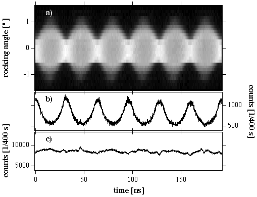 |
| Figure 1: Intensity distribution in the
rocking-angle time coordinate plane, a). Nodes and
anti-nodes of the standing waves are distinguished
clearly. b) shows a section through the center of the
rocking curve having maximal intensity when the rocking
curve is narrowest while the integrated intensity, c),
remains constant as a function of time,. The latter does
not depend on the momentary strain state and indicates
that the diffraction is kinematic. The excitation
frequency is 15.5991 MHz. |
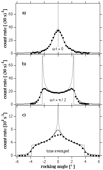 |
| Figure 2: Snap shots for an excited crystal at moments where the lattice is mostly relaxed and at maximum strain given by the large dots in a) and b), respectively. Plot c) gives time averaged data obtained without any timing setup. All three curves follow well the behavior predicted by the theory, a Dirac delta function in a), an inverse circle function in b) and an complete elliptic integral of the first kind in c). The theoretical distributions are given by the dotted lines whereas the continuos lines in b) and c) are folded with the same Lorentzian resolution function. |