THE CRYSTAL STRUCTURE OF A CYCLOAMYLOSE WITH 26 GLUCOSE RESIDUES AND THE NEW MODEL FOR A 6-FOLD V-AMYLOSE
Katrin Gessler1, Isabel Uson2, Takeshi Takaha3, Norbert Krauss1, Steven Smith4, George Sheldrick2, Wolfram Saenger1
1Institut für Kristallographie, Freie
Universität Berlin, Takustrasse 6, 14195 Berlin, Germany; 2Institut
für anorganische Chemie, Universität Göttingen, Tammanstrasse
4, 37077 Göttingen, Germany;
3Biochemical Research Laboratory,
Ezaki Glico Co., Ltd., 4-5-6 Utajima, Nishiyodogawa, Osaka 555,
Japan;
4Insitute of Cell and Molecular
Biology, University of Edinburgh, The kings buildings, Mayfield
Road, Edinburgh EH9 3JH, United Kingdom
Cycloamyloses (CA) are cyclic compounds consisting of a-1,4-linked D-glucose residues and can be obtained by enzymatic treatment of synthetic amylose with CGTases. The smallest members of this family are the a,b and g-cyclodextrins (CA6, 7 and 8) which contain 6,7 and 8 glucose residues, respectively. Recently, the reaction of a D-enzyme could be established and yields larger cycles up to a dp of several 100 glucoses [1]. NMR investigations have shown that there are changes in the chemical shift of the carbons C1 and C4 (those carbons which are involved in the glycosidic bond) for all cycloamyloses larger than 9 residues, indicating structural changes, which could be confirmed in the crystal structures of CA10 and CA14: in these compounds two new structural motifs could be observed; the 'kink' and the 'flip' [2]. What we have called 'flip' is a 180o rotation of a glucose residue with respect to its neighbour leading to elliptical structures with narrow, groove-like cavities.
As expected from the NMR-data, 'flip'-sites could also be observed in the crystal structure of a larger cycloamylose with 26 glucose residues.
This compound crystallizes in a triclinic space group, P1, with two CA26 molecules and 76.8 cocrystallized water molecules in the asymmetric unit; an overall content of 648.6 non-hydrogen atoms. The structure has been solved 'ab initio' (program SHELXD) and is up to now the largest unknown light atom structure being solved by direct methods. The refinement (program SHELXL) with high resolution data up to 0.99Å resolution collected at synchrotron (beamlines EMBL at DESY, Hamburg and LURE, Orsay) converged to a final R-value of 0.082 (Rfree 0.101).
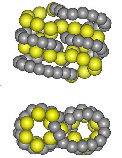
The two CA26 molecules in the asymmetric unit have a similar overall shape: Each of them is composed of two left-handed 6-fold helices connected by two flips in an antiparallel arrangement. The hydrophobic central cavity of the helix and the intersticies are filled with the 76.8 cocrystallized water molecules distributed over 147 positions. A coordinate set for a 6-fold amylose helix with a height of 7.8Å and diameter (inner 5Å, outer 14.5Å) similar to a-cyclodextrin, could be derived from the central glucoses of CA26. Intramolecular hydrogen bonds O2(n)...O3(n-1) and O6(n)...O3(n+1) stabilize the helix and interstitial water molecules connect neighboured helices by short hydrogen bonds.
The crystal structure of CA26 gives not only a good explanation for the hydratation of a V-amylose helix, but also a model for the chain folding of polymer helices, since the introduced flips allow a very tight and low energy packing of amylose chains.
Fig: For clarity, every glucose has been reduced to 3 atoms: C6 (big balls) and O2 and O3 (smaller balls). Top: side-view, bottom: top-view. The CA26 molecule is clearly divided into two parts connected by the flip sites. Each part consists of 13 glucose residues and forms two turns of a left-handed single helix.
1: Terada, Y., Yanase, M., Takata, H., Takaha, T., Okada, S., J.Biol.Chem.,
272, 15729-15733, (1997)
2: Jacob, J., Gessler, K., Hoffmann, D., Sanbe, H., Koizumi, k.,
Smith,S., Takaha, T., Saenger, W., Angew.Chem., 37,
605-609, (1998)}