
HIGH RESOLUTION X-RAY DIFFRACTION INVESTIGATION OF MBE-GROWN QUASI-HOMOEPITAXIAL 3C-SiC LAYERS ON 6H-SiC SUBSTRATES
Kräusslich, J., Köcher, B., Bauer, A. and Goetz, K.1, Fissel, A. and Richter,W.2
1 Institute of Optics and
Quantumelectronics, Friedrich Schiller University Jena,
Max-Wien-Platz 1, D-07743 Jena, phone: +49-3641-947254, fax:
+49-3641-947202, e-mail: onk@uni-jena.de
2 Institute of Solid State Physics, Friedrich
Schiller University Jena
3C-SiC thin films have been investigated by means of high resolution X-ray diffraction techniques (w /2Q-scan, Q-scan, n-scan) and X-ray topography. The SiC films were grown in a solid source molecular beam epitaxy (MBE) system on 6H-SiC substrate crystals at depositions temperatures in the range from 780°C to 950oC and growth rates in the range from 30 nm/h to 120 nm/h.
The symmetric high resolution X-ray diffractogram shows two clearly separated family reflections (6H-SiC: 00.12; 3C-SiC: 222), which have their origin in the 6H-SiC substrate and SiC thin film, respectively. This indicates a significant difference of the c/q lattice parameter (q - number of Si-C double layers per unit cell) in the order of 2.19·10-4 nm (Fig.1).
FIG. 1: High resolution X-ray diffractogram of MBE deposited 3C-SiC thin film on 6H-SiC substrate. w -2J -scan of the symmetric 6H-SiC 00.12 and the 3C-SiC 222 reflection, respectively.
We also investigated the lattice mismatch parallel and perpendicular to the [00.1]-direction. For this we used the so called “reciprocal space mapping” technique. Rocking curves were measured at several fixed 2J -positions. We got a two-dimensional image of the reciprocal space close to a reciprocal lattice point (Fig 2). In the case of 3C-SiC on 6H-SiC the epilayer had crystallographic pseudomorph structure (lateral lattice fit, normal allowed lattice difference) i.e. there is almost no lateral lattice mismatch. The mismatch parallel to the c-axis D c/c was 8.7·10-4.
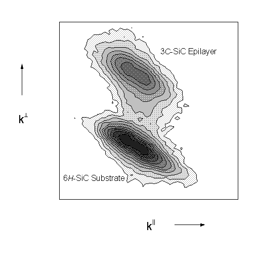
FIG. 2: Reciprocal space mapping of the asymmetric 11.12 reflection of a 1.2 µm MBE grown 3C-SiC layer on a 6H-SiC Substrate.
When 3C-SiC films were grown on 6H-SiC substrates, double position boundaries (stacking sequence ACB instead of ABC) were frequently observed. For the visualization of this effect high resolution X-ray topographic measurements are investigated at 3C-SiC layers on 6H-SiC substrates. We find, that the domains of different stacking sequences are equally distributed in the 3C-SiC epilayer. The size of this domains are approximately 200 × 200 µm2.