STRUCTURE OF HUMAN LUNG MAST CELL b-TRYPTASE, A SERINE PROTEINASE INVOLVED IN ALLERGIC ASTHMA
Pedro Jose Barbosa Pereira1,
Andreas Bergner1, Sandra Macedo-Ribeiro1,
Robert Huber1, Gabriele Matschiner2,
Hans Fritz2, Christian P. Sommerhoff2
and Wolfram Bode1
1 Abt. für
Strukturforschung, Max-Planck-Institut für Biochemie,
Martinsried, Germany
2 Abt. für Klinische
Chemie und Klinische Biochemie in der Chirurgischen Klinik und
Poliklinik, Klinikum Innenstadt der LMU, München, Germany
Keywords: asthma, tryptase, serine proteinase
Human tryptase (EC 3.4.21.59), the predominant protein of most human mast cells, has been implicated in the pathogenesis of asthma and other allergic and inflammatory disorders[1].
Although tryptase displays striking similarities with other serine proteinases, it has a number of unique properties (reviewed in refs. 1,2):
Tryptase efficiently hydrolyses a number of (neuro-)peptide substrates (in vitro) in a trypsin-like manner[3]. Unlike trypsin, however, tryptase cleaves only a few proteins, among these fibrinogen, fibronectin and high molecular weight kininogen (inactivation), and the zymogens of stromelysin-1 and u-PA (activation).
Attempts to model the structure of tryptase[4-6] are necessarily based on monomeric serine proteinases, and are thus unable to predict the tetrameric architecture. In order to define the tryptase-tetramer and to obtain a reliable model for rational drug design, we determined the X-ray crystal structure of human lung mast cell b-tryptase.
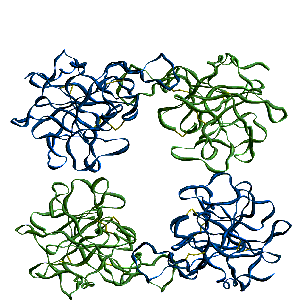
The 3 A crystal structure of human b-tryptase[7]
in complex with 4-amidinophenylpyruvic acid reveals four
quasi-equivalent monomers arranged in a square flat ring. The
four active centres of the tetramer are directed towards an oval
central pore, restricting the access for macromolecular
substrates and inhibitors. Heparin chains could stabilise the
complex by binding to an elongated patch of positively charged
residues spanning two adjacent monomers.
Fig. 1: Overall architecture of the
tryptase tetramer (front view). The four protease monomers are
disposed in the corners of a square, with active sites facing the
central pore.
This unique tetrameric architecture explains many of
tryptaseís distinct biochemical properties and provides a basis
for the rational design of mono- and multifunctional tryptase
inhibitors.
Acknowledgements: We thank D. Grosse for
her excellent help in crystallisation. This work was supported by
scholarships PRAXIS XXI/BD/9782/96 (to P. J. B. P.) and PRAXIS
XXI/BD/4050/94 (to S. M-R.) from the Fundaçäo para a Ciencia e
a Tecnologia, Portugal, and by Biotech programs of the European
Union, by the Sonderforschungsbereich 469 of the University of
Munich, by the Deutsche Forschungsgemeinschaft and by the Fonds
der Chemischen Industrie. P. J. B. P. is a Programa Gulbenkian de
Doutoramento em Biologia e Medicina fellow.